Session Information
Date: Sunday, November 7, 2021
Title: Pediatric Rheumatology – Clinical Poster II: SLE, JDM, & Juvenile Scleroderma (0764–0785)
Session Type: Poster Session B
Session Time: 8:30AM-10:30AM
Background/Purpose: Craniofacial scleroderma (Cf-LS), also known as Parry-Romberg Syndrome or scleroderma en coup de sabre, is a subtype of localized scleroderma (morphea). Diagnosis and monitoring of Cf-LS is challenging due to the spectrum of clinical manifestations and lack of robust standardized assessment tools. Digital 3D facial maps help to provide objective assessment of longitudinal facial tissue changes between clinical visits. Traditional stationary camera systems are large, expensive, and require specialized training to use. In contrast, handheld 3D cameras are portable, low-cost, and easy to use. Handheld 3D cameras have been shown to produce reliably high-quality 3D facial maps in adults both with and without craniofacial differences. We set out to determine if the same holds true for children with Cf-LS.
Methods: Digital facial images were obtained on 22 clinically stable Cf-LS patients < 23 years of age (IRB #19080205). Each subject was photographed with a handheld Vectra H2 (Canfield Scientific) and stationary 3dMD.head.t camera systems (3dMD), with a randomized order of acquisition. Number of trials and acquisition time to obtain a satisfactory image were recorded. Descriptive statistics were reported. Consensus facial landmarks (FaceBase Consortium) were digitally applied to each map by two trained individuals. Linear and curvilinear distances between landmarks were measured (VAM software). Reliability statistics and magnitude of error between instruments were calculated.
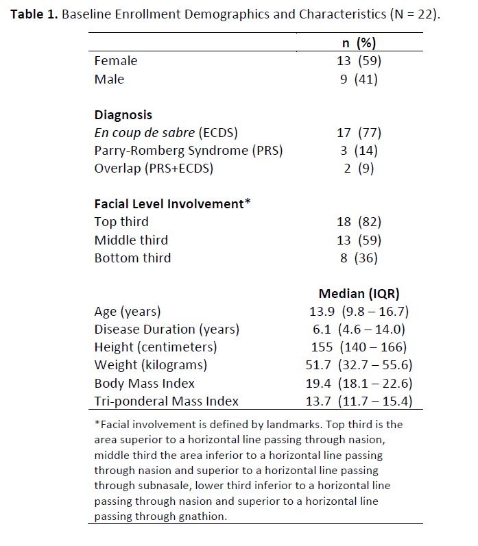
Results: Subjects ranged in age from 3.5 – 21.5 years with a median disease duration of 6.1 years (Table 1). Mean total image acquisition time was not significantly different between the 3dMD and Vectra camera systems (3dMD = 59 seconds vs. Vectra = 61 seconds, p=0.71). Age was poorly related to acquisition time for both Vectra (Pearson r = -0.07) and 3dMD (Pearson r = -0.32) systems. Height was poorly related to acquisition time for the Vectra system (Pearson r = -0.21) but moderately related for the 3dMD system (Pearson r = -0.41).
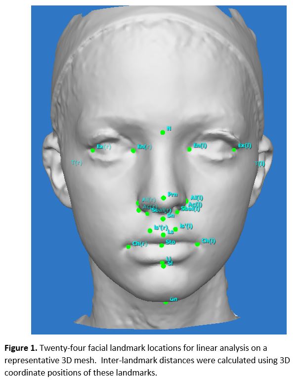
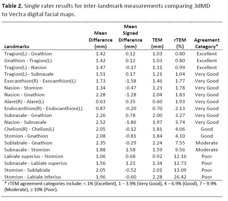
Comparing the digital facial maps (Figure 1) from 3dMD and Vectra across 19 inter-landmark linear distances and 2 raters, the median technical error of measurement (TEM) was 1.91mm (range 0.6 – 9.08mm) and the median relative TEM (rTEM) was 3.85% (range 0.80% – 26.42%) (Table 2). Measurements with the highest TEM and rTEM values usually involved oral and/or peri-oral landmarks. Only 36% of subjects had Cf-LS involvement of the lower third of the face. Measurements with the lowest TEM and rTEM values typically included landmarks along the midline of the face.
Conclusion: Handheld 3D cameras are easy to use and generate high quality digital facial maps in children as young as 3 years of age with craniofacial scleroderma, which are overall equivalent to images obtained by stationary cameras. Age and height did not impact the ease of obtaining handheld camera images. Measurements utilizing oral and peri-oral landmarks are most vulnerable to variation due to subtle changes in facial expression between acquisition on both camera systems. Future work will explore the accuracy of longitudinal digital facial maps for disease monitoring in the same population.
To cite this abstract in AMA style:
Glaser D, Schollaert-Fitch K, Liu C, Goldstein J, Torok K. Pediatric Craniofacial Scleroderma: Assessing Handheld 3D Stereophotogrammetric Imaging Feasibility and Reliability [abstract]. Arthritis Rheumatol. 2021; 73 (suppl 9). https://acrabstracts.org/abstract/pediatric-craniofacial-scleroderma-assessing-handheld-3d-stereophotogrammetric-imaging-feasibility-and-reliability/. Accessed .« Back to ACR Convergence 2021
ACR Meeting Abstracts - https://acrabstracts.org/abstract/pediatric-craniofacial-scleroderma-assessing-handheld-3d-stereophotogrammetric-imaging-feasibility-and-reliability/


