Session Information
Date: Sunday, November 12, 2023
Title: (0066–0095) T Cell Biology & Targets in Autoimmune & Inflammatory Disease Poster
Session Type: Poster Session A
Session Time: 9:00AM-11:00AM
Background/Purpose: Programmed death 1 (PD-1) is an immune checkpoint receptor expressed by activated T cells, and of major importance in maintaining peripheral tolerance. Expression of PD-1 has been associated with T cells exhaustion, plasma cell differentiation and bone homeostasis. With emerging treatments engaging the PD-1 pathway in Rheumatoid Arthritis (RA) this pathway is of continued interest.
We aimed to investigate PD-1hi T cells from RA synovial fluid (SF).
Methods: SF was collected from RA patients with disease flare (n=7). Cells were stained for flow and sorted in CD3+ PD-1hi/PD-1lo populations. Post sorting, bulk RNA seq was performed, and cells were stimulated with antiCD3/CD28. Cytokines in the supernatants were investigated using V-plex multipanel assay. Supernatants were added to fibroblast cultures and the osteoblast cell line SaOs-2.
Results: Evaluated by flow cytometry, PD-1+ cells represented 42% of CD4+ T cells and 34% of CD8+ T cells in the SF. When comparing co-expression of checkpoint receptors and transcription factors between PD-1hi and PD-1lo CD4+ T cell; Tigit, Tim3 and NFAT were all increased in PD-1hi (all p < 0.05), Lag-3 was slightly elevated (p=0.07), whereas TCF1 and TOX expression did not differ. These differences were not observed between PD-1hi and PD-1lo CD8+ T cells.
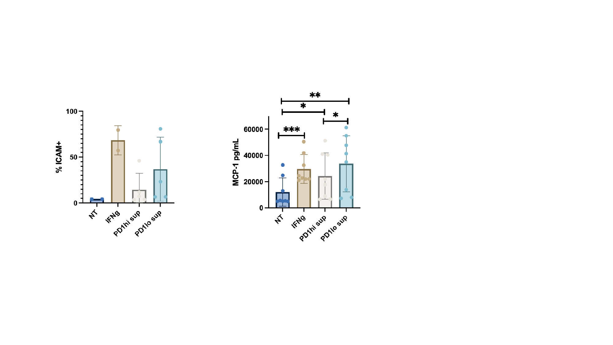
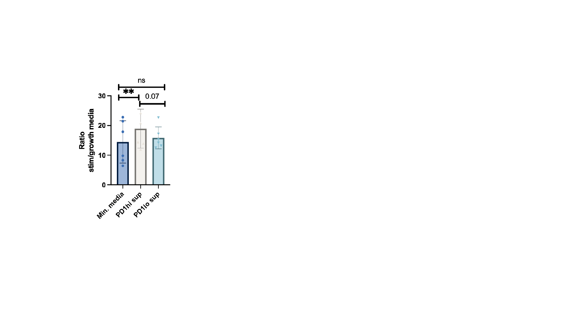
Post sorting, RNAseq revealed PD-1hi and PD-1lo cells to cluster separately. In PD-1hi cells, gene expression corresponded to structural signatures, including tubulin binding and cytoskeletal motor activity (p < 0.01). Genes related to peripheral T helper cells, including IL21 and CXCL13 were highly upregulated (all p< 0.05) The gene PHEX was highly expressed (log2 fold change=5.2), suggesting an association with bone mineralization. The signature in PD-1lo cells was associated with immune activation genes (p < 0.005). Stimulating cells with CD3/CD28 post sorting resulted in lower production of IL-2, IFNg, TNFa, IL-8, IL-10, IL-13, IL-12, and IL-1b from PD-1hi cells compared to PD-1lo. The supernatant from PD-1hi cells only slightly activated fibroblasts, whereas the PD-1lo supernatant significantly activated fibroblasts, as measured by by MCP-1 production and surface ICAM expression (fig 1). When adding the PD-1hi and PD-1lo supernatant to osteoblast cultures, bone mineralization was significantly increased by the PD-1hi supernatant (fig 2).
Conclusion: PD-1hi cells are present in the synovial joint. They are not exhausted but express multiple checkpoint receptors, suggesting regulatory capacities. They could play a central role in structural maintenance through effects on fibroblasts and osteoblasts.
To cite this abstract in AMA style:
Rask S, Andersen C, Hvid M, Ravnskjær K, Deleuran B, Greisen S. PD-1hi Cells Are Highly Present in the Rheumatoid Joint, and Express Regulatory Capacities [abstract]. Arthritis Rheumatol. 2023; 75 (suppl 9). https://acrabstracts.org/abstract/pd-1hi-cells-are-highly-present-in-the-rheumatoid-joint-and-express-regulatory-capacities/. Accessed .« Back to ACR Convergence 2023
ACR Meeting Abstracts - https://acrabstracts.org/abstract/pd-1hi-cells-are-highly-present-in-the-rheumatoid-joint-and-express-regulatory-capacities/


