ACR Meeting Abstracts

ACR Meeting Abstracts

  • Meetings
    • ACR Convergence 2024
    • ACR Convergence 2023
    • 2023 ACR/ARP PRSYM
    • ACR Convergence 2022
    • ACR Convergence 2021
    • ACR Convergence 2020
    • 2020 ACR/ARP PRSYM
    • 2019 ACR/ARP Annual Meeting
    • 2018-2009 Meetings
    • Download Abstracts
  • Keyword Index
  • Advanced Search
  • Your Favorites
    • Favorites
    • Login
    • View and print all favorites
    • Clear all your favorites
  • ACR Meetings

Abstract Number: 0256

Patients with ACPA-positive and ACPA-negative Rheumatoid Arthritis Show Different Circulating Auto-antibody Repertoires

Kevin Cunningham1, Benjamin Hur2, John Davis2 and Jaeyun Sung2, 1University of Minnesota, Rochester, MN, 2Mayo Clinic, Rochester, MN

Meeting: ACR Convergence 2022

Keywords: Anti-citrullinated Protein Autoantibodies (ACPAs), autoantigens, Bioinformatics, Biomarkers, rheumatoid arthritis

  • Tweet
  • Click to email a link to a friend (Opens in new window) Email
  • Click to print (Opens in new window) Print
Session Information

Date: Saturday, November 12, 2022

Title: RA – Diagnosis, Manifestations, and Outcomes Poster I

Session Type: Poster Session A

Session Time: 1:00PM-3:00PM

Background/Purpose: RA is commonly diagnosed through a serological test for the presence of ACPA, and RA patients who test positive are collectively known as ‘ACPA-positive RA’ (ACPA+). However, patients can test negative for ACPA yet still be clinically diagnosed with RA, and are thereby designated as ‘ACPA-negative RA’ (ACPA–). Although ACPA+ and ACPA– patients are known to have different rates of erosive damage and distinct risk factors, the differences in serological profiles between these two disease subgroups are largely unknown. In this study, we performed a comprehensive survey of serum autoantibodies in patients with ACPA+ and ACPA– RA to obtain novel insights into autoantigens underlying the distinct subgroups of RA.

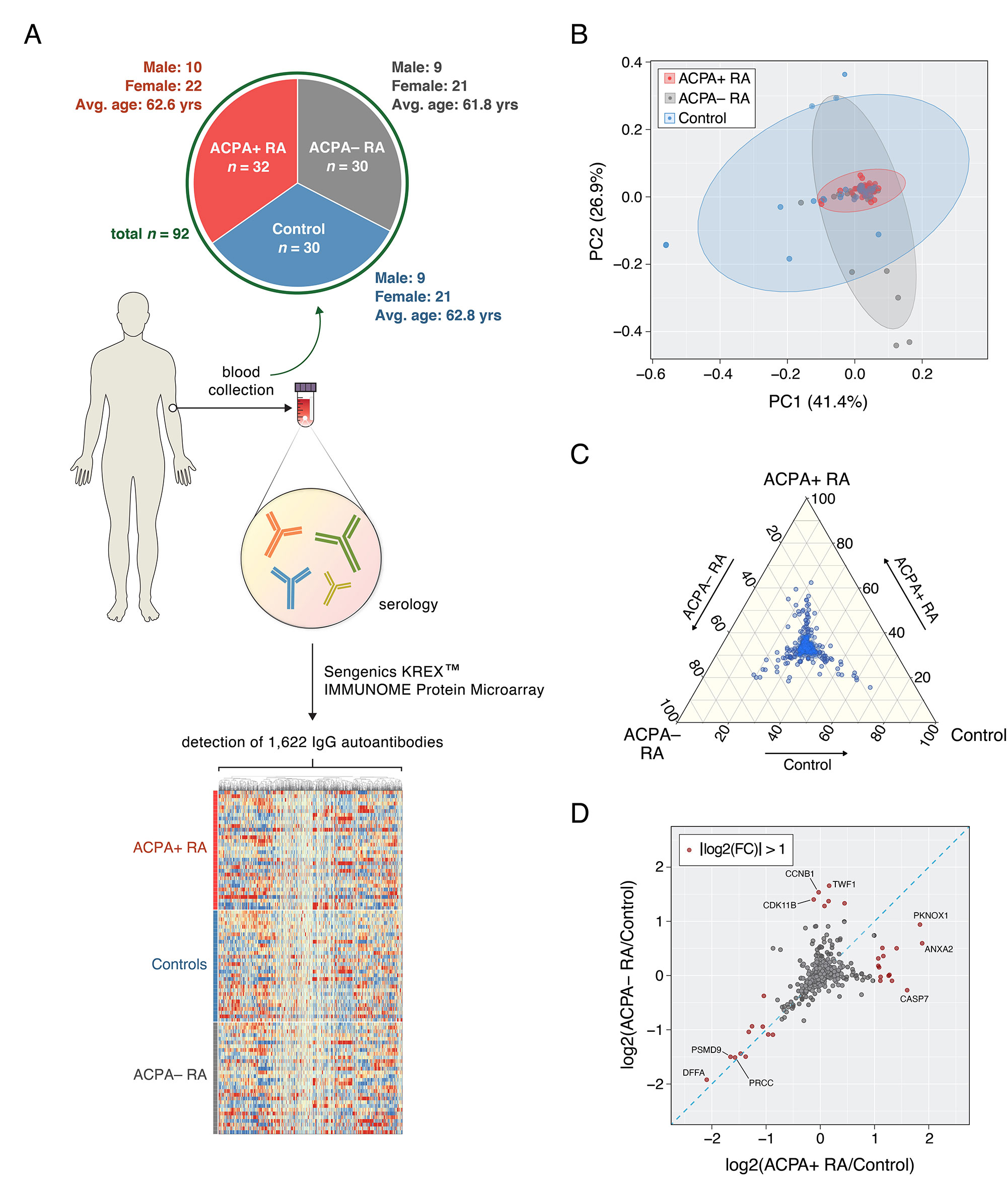
Methods: Serum was collected from patients with ACPA+ RA (n = 32), patients with ACPA– RA (n = 30), and healthy controls (n = 30) (Fig. 1A). Sengenics KREXTM multiplexed autoantibody-screening protein microarray was used to screen for 1,622 different autoantibodies from each serum sample. A Mann–Whitney U test combined with Cliff’s delta was used to identify differentially abundant autoantibodies.

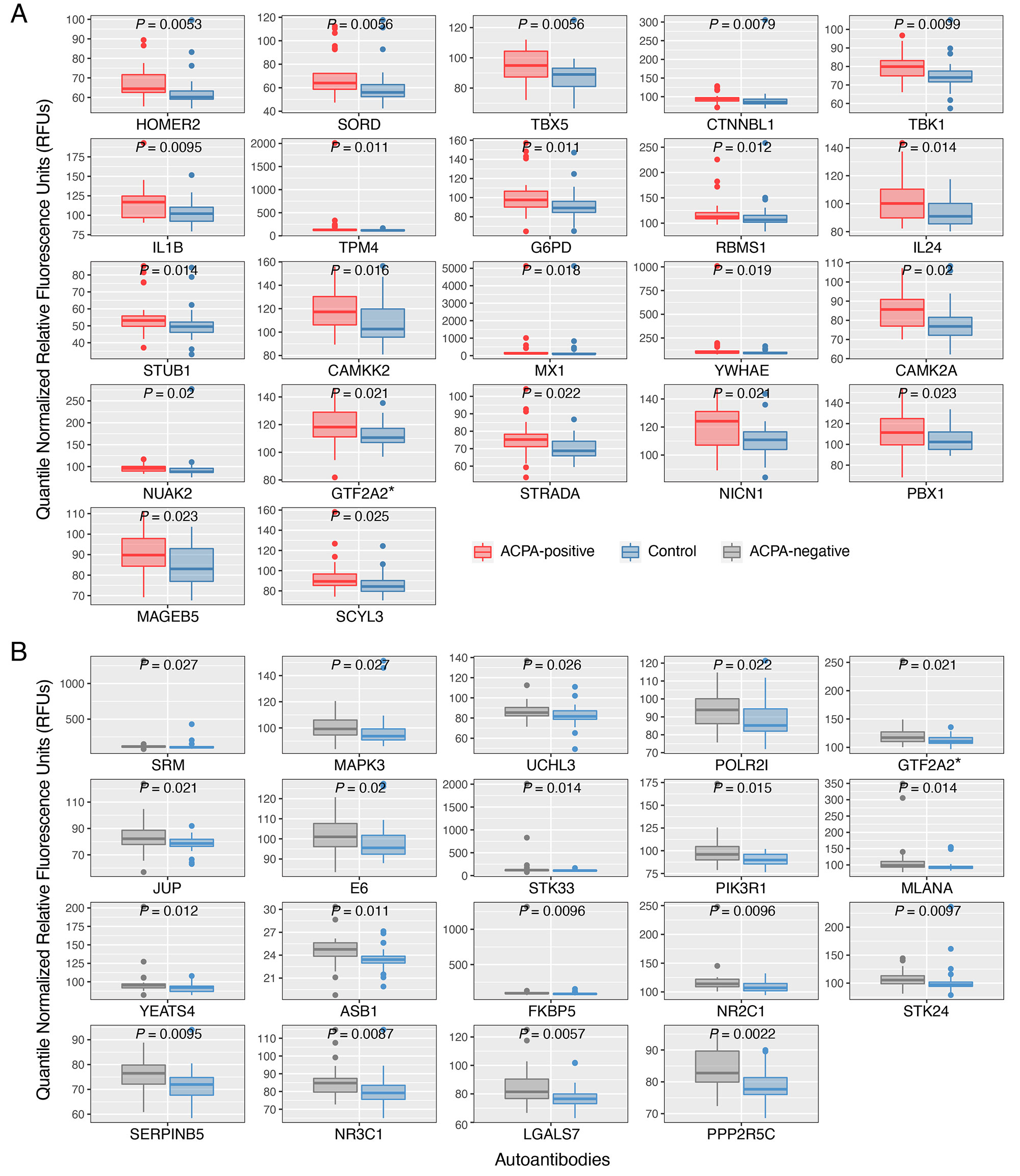
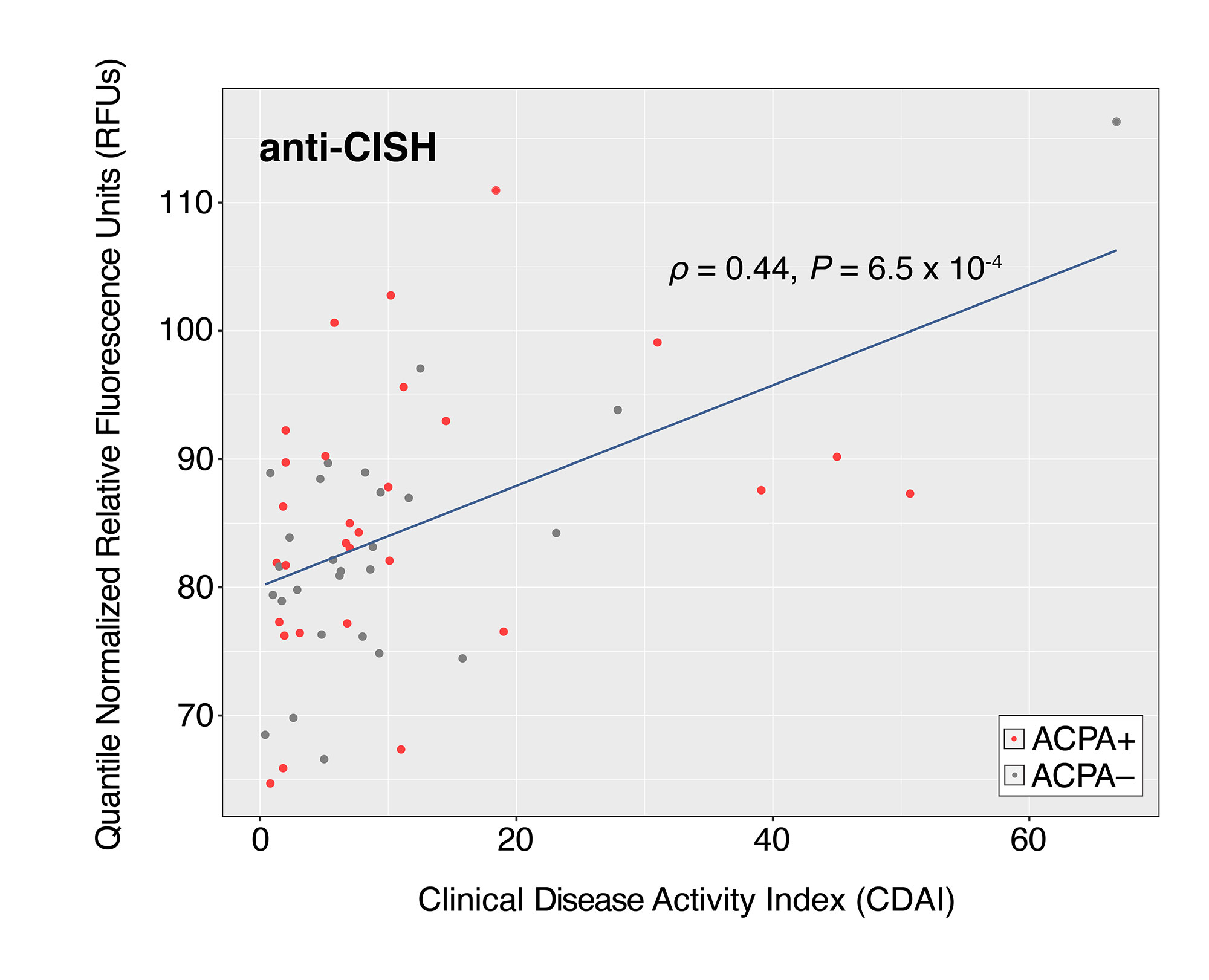
Results: ACPA+ RA patients, ACPA– RA patients, and healthy controls displayed differences in their serum autoantibody profiles (Fig. 1B–D). Upon closer examination, we identified differential serum autoantibodies of ACPA+ and ACPA– RA. Specifically, we found 22 and 19 autoantibodies higher in ACPA+ RA patients and ACPA– RA patients, respectively, compared to healthy controls (Fig. 2A–B). Notably, only one autoantibody for GTF2A2 was found to be higher in both RA subgroups. Next, we found autoantibodies for the CISH protein to be positively correlated with the Clinical Disease Activity Index (ρ = 0.44, P = 6.5 × 10⁻⁴) (Fig. 3). Additionally, we determined the functional classes of the proteins targeted by the differentially abundant autoantibodies. We found eight different functional classes, using the PANTHER classification system, for the autoantibodies higher in ACPA+ RA patients, and seven different functional classes for the autoantibodies higher in ACPA– RA patients. When comparing these two groups of functional classes, we identified four in common: ‘Gene-specific Transcriptional Regulator’, ‘Nucleic Acid Metabolism Protein’, ‘Metabolite Interconversion Enzyme’, and ‘Protein-modifying Enzyme’. This suggests that an imbalance in autoantibody production targeting metabolic and transcription processes could potentially be a newly identified hallmark of RA.

Conclusion: We identified several differentially abundant autoantibodies in ACPA+ and ACPA– RA, many of which were identified for the first time. Our study demonstrates the potential utility of serum autoantibodies as diagnostic targets for RA subgroups and reveals potential therapeutic treatments.

Supporting image 1

Figure 1. Study design overview and group-wise comparisons of autoantibody composition profiles. (A) Blood (serum) samples were collected to investigate serum autoantibody compositions in ACPA+ RA (n = 32), ACPA– RA (n = 30), and healthy controls (n = 30). Each serum sample was screened for 1,622 IgG autoantibodies using the Sengenics KREXTM IMMUNOME Protein Microarray. Raw relative fluorescence units (RFU) values representing serum autoantibody abundances were transformed using quantile normalization prior to all downstream analyses. Heatmap illustrates autoantibodies clustered according to abundance similarities across samples. (B) Principal component analysis on the 92 autoantibody profiles from ACPA+, ACPA–, and healthy controls. Controls showed the highest within-group heterogeneity in autoantibody composition whereas the ACPA+ RA group showed the least within-group heterogeneity. (C) Ternary plot showing mean abundances of 1,622 autoantibodies across ACPA+, ACPA–, and healthy controls. Coordinates of each point correspond to percentages of mean abundances and sum to 100. Despite most autoantibodies having similar mean abundances across the three study groups, several autoantibodies were found to have high abundance in one group (see points closer to the corners). (D) Fold-changes in mean abundances of 1,622 autoantibodies for the RA subgroups compared to the control group. x-axis and y-axis correspond to the fold-changes between ACPA+ RA and controls and between ACPA– RA and controls, respectively. Autoantibodies shown in red points have a fold-change of 2 (or greater) between groups. As observed in (C), several autoantibodies show fold-changes specific to an RA subgroup. The blue diagonal dashed line represents the line y=x.

Supporting image 2

Figure 2. Autoantibodies identified to have significantly higher abundances in two subgroups of rheumatoid arthritis (ACPA+ RA and ACPA– RA) than in healthy controls. Patients with (A) ACPA+ RA and (B) ACPA– RA show higher abundances in 22 and 19 autoantibodies, respectively. Two-sided Mann–Whitney U test (P < 0.05) and Cliff’s Delta (|d| > 0.33) were used to find autoantibodies of significantly higher abundance. Standard box-and-whisker plots (e.g., center line, median; box limits, upper and lower quartiles; whiskers, 1.5× interquartile range; points, outliers) are used to show numerical data. *, auto-antibody for GTF2A2 protein was found to have significantly higher abundances in both ACPA+ and ACPA– RA patient groups than in healthy controls.

Supporting image 3

Figure 3. Autoantibodies targeting CISH proteins display the highest correlation with Clinical Disease Activity Index (CDAI). The blue line in the scatterplots represents the linear regression fit of Y~X, wherein X and Y correspond to CDAI and autoantibody abundance, respectively. The Spearman correlation coefficient ρ was used to measure the strength of the relationship between the two variables. Scatterplot contains 56 points corresponding to the number of RA patients whose CDAI scores were available.


Disclosures: K. Cunningham, None; B. Hur, None; J. Davis, Pfizer; J. Sung, None.

To cite this abstract in AMA style:

Cunningham K, Hur B, Davis J, Sung J. Patients with ACPA-positive and ACPA-negative Rheumatoid Arthritis Show Different Circulating Auto-antibody Repertoires [abstract]. Arthritis Rheumatol. 2022; 74 (suppl 9). https://acrabstracts.org/abstract/patients-with-acpa-positive-and-acpa-negative-rheumatoid-arthritis-show-different-circulating-auto-antibody-repertoires/. Accessed .
  • Tweet
  • Click to email a link to a friend (Opens in new window) Email
  • Click to print (Opens in new window) Print

« Back to ACR Convergence 2022

ACR Meeting Abstracts - https://acrabstracts.org/abstract/patients-with-acpa-positive-and-acpa-negative-rheumatoid-arthritis-show-different-circulating-auto-antibody-repertoires/

Advanced Search

Your Favorites

You can save and print a list of your favorite abstracts during your browser session by clicking the “Favorite” button at the bottom of any abstract. View your favorites »

All abstracts accepted to ACR Convergence are under media embargo once the ACR has notified presenters of their abstract’s acceptance. They may be presented at other meetings or published as manuscripts after this time but should not be discussed in non-scholarly venues or outlets. The following embargo policies are strictly enforced by the ACR.

Accepted abstracts are made available to the public online in advance of the meeting and are published in a special online supplement of our scientific journal, Arthritis & Rheumatology. Information contained in those abstracts may not be released until the abstracts appear online. In an exception to the media embargo, academic institutions, private organizations, and companies with products whose value may be influenced by information contained in an abstract may issue a press release to coincide with the availability of an ACR abstract on the ACR website. However, the ACR continues to require that information that goes beyond that contained in the abstract (e.g., discussion of the abstract done as part of editorial news coverage) is under media embargo until 10:00 AM ET on November 14, 2024. Journalists with access to embargoed information cannot release articles or editorial news coverage before this time. Editorial news coverage is considered original articles/videos developed by employed journalists to report facts, commentary, and subject matter expert quotes in a narrative form using a variety of sources (e.g., research, announcements, press releases, events, etc.).

Violation of this policy may result in the abstract being withdrawn from the meeting and other measures deemed appropriate. Authors are responsible for notifying colleagues, institutions, communications firms, and all other stakeholders related to the development or promotion of the abstract about this policy. If you have questions about the ACR abstract embargo policy, please contact ACR abstracts staff at [email protected].

Wiley

  • Online Journal
  • Privacy Policy
  • Permissions Policies
  • Cookie Preferences

© Copyright 2025 American College of Rheumatology