ACR Meeting Abstracts

ACR Meeting Abstracts

  • Meetings
    • ACR Convergence 2024
    • ACR Convergence 2023
    • 2023 ACR/ARP PRSYM
    • ACR Convergence 2022
    • ACR Convergence 2021
    • ACR Convergence 2020
    • 2020 ACR/ARP PRSYM
    • 2019 ACR/ARP Annual Meeting
    • 2018-2009 Meetings
    • Download Abstracts
  • Keyword Index
  • Advanced Search
  • Your Favorites
    • Favorites
    • Login
    • View and print all favorites
    • Clear all your favorites
  • ACR Meetings

Abstract Number: 0090

Parenchymal and Hematopoietic IFNγ Signaling Exhibits Dichotomous Effects in Murine Lupus

Minjung Kim1, Anthony Marinov2, Haylee Cosgrove3, Xiaoyan Gong3, Mark Shlomchik3 and Jeremy Tilstra3, 1University of Illinois College of Medicine at Chicago, Chicago, IL, 2University of Pittsburgh, Homestead, PA, 3University of Pittsburgh, Pittsburgh, PA

Meeting: ACR Convergence 2024

Keywords: Animal Model, cytokines, Lupus nephritis, Systemic lupus erythematosus (SLE)

  • Tweet
  • Click to email a link to a friend (Opens in new window) Email
  • Click to print (Opens in new window) Print
Session Information

Date: Saturday, November 16, 2024

Title: SLE – Animal Models Poster

Session Type: Poster Session A

Session Time: 10:30AM-12:30PM

Background/Purpose: Lupus nephritis is the most common life-threatening end-organ complication of SLE, and response rates to current interventions remain poor. Notably, PD-L1 and other interferon-responsive genes are upregulated in the kidneys of both humans and mice with lupus nephritis. We and others have shown that the kidney parenchyma has immunosuppressive effects on infiltrating immune cells. We hypothesize that the kidney parenchyma upregulates several factors in response to inflammatory signals, which can then dampen inflammatory signals to reduce local damage in the kidney. As IFNγ is the major inducer of PD-L1, we postulated that IFNγ induces this protective program in the kidneys of lupus-prone mice.

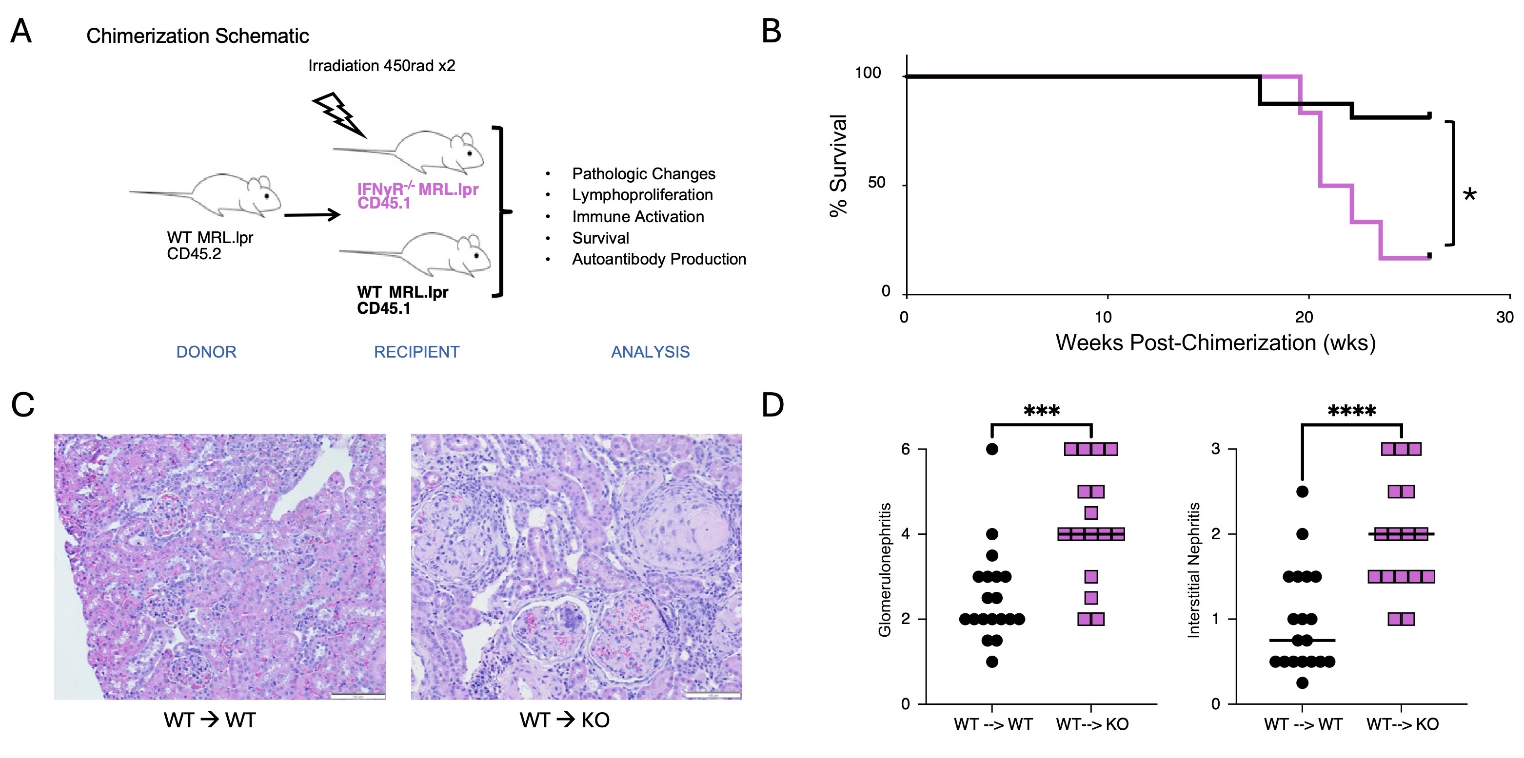
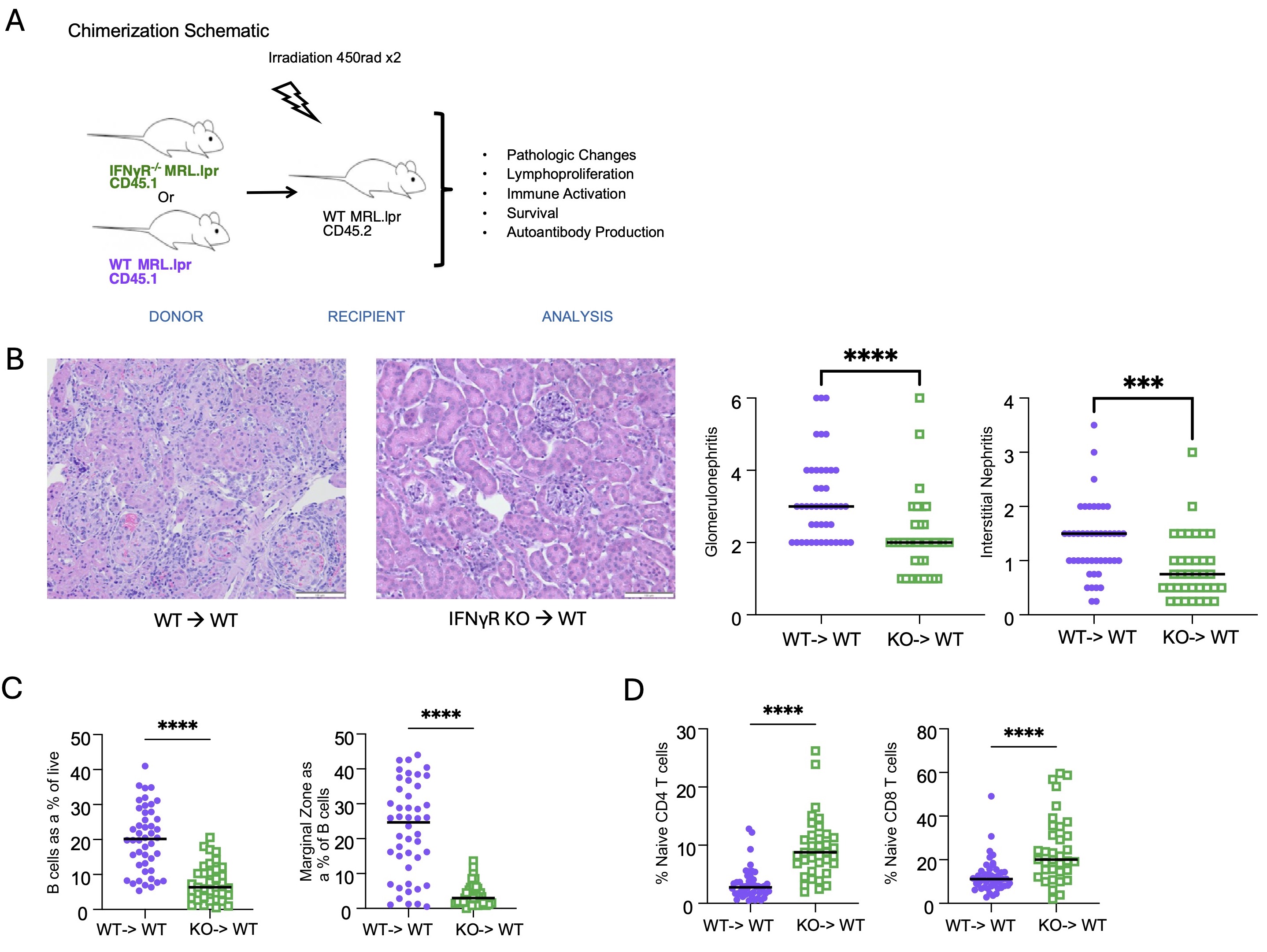
Methods: MRL.Faslpr mice develop autoantibodies, proteinuria, dermatitis, and glomerulonephritis. In this study, we created parenchymal bone marrow chimeras in which congenically labeled wild-type (WT) immune cells are transferred into either WT MRL.Faslpr, IFNγR-/- MRL.Faslpr, or PD-L1-/- MRL.Faslpr recipients (Fig 1A). Based on our hypothesis, we predicted that both the IFNγR-/- and PD-L1-/- recipients would have more severe disease than WT controls. We also performed reverse chimeras in which WT or IFNγR-/- bone marrow was transferred into WT MRL.Faslpr recipients to assess the role of IFN signaling in the hematopoietic compartment (Fig 2A). Analysis included measurement of proteinuria, histological assessment of interstitial and glomerular disease, autoantibody quantification, FACS analysis of immune cell activation, and survival analysis.

Results: Consistent with our hypothesis, IFNγR-/- MRL.Faslpr recipient mice demonstrated more severe and rapid disease onset than WT recipient controls; this included higher rates of mortality (p< 0.05), more severe glomerulonephritis (p< 0.005), and interstitial disease (p< 0.001) (Fig 1). While parenchymal IFNγR deficiency resulted in reduced PD-L1 expression, unexpectedly, the PD-L1-/- MRL.Faslpr recipient mice did not exhibit an exacerbated phenotype, suggesting an alternative regulatory mechanism induced by IFNγ in the kidney of lupus-prone mice. Conversely, when IFNγR was deleted in the hematopoietic compartment, mice exhibited ameliorated disease (Fig 2).

Conclusion: Immune signaling mediated by IFNγ has dichotomous effects in lupus-prone mice. In the parenchyma, IFNγ regulates an anti-inflammatory pathway that surprisingly is independent of PD-L1, while in the hematopoietic compartment, IFNγ has pro-inflammatory properties. These findings are key to informing future therapeutic development as inhibiting receptors or inflammatory signaling pathways on specific cell types may be more efficacious than targeting these pathways systemically.  This may explain why anifrolumab, which is efficacious for systemic disease, has had suboptimal responses in lupus nephritis trials as it may lead to proinflammatory effects in the kidney. These findings may also aid our understanding of the fundamental biology of disease; for instance, they demonstrate that the kidney is not an innocent bystander, but can play a role in regulating inflammation, which could explain underlying tissue-specific disease heterogeneity.

Supporting image 1

Figure 1: Schematic of bone marrow chimerization for IFNγR-/- or WT recipients receiving congenically marked CD45.2 MRL.lpr bone marrow (A) IFNγR-/- recipients exhibit increased mortality (B), which is associated with accelerated nephritis as noted by representative histology (C) and histologic analysis for glomerular disease and interstitial nephritis (D). With pink lines and dots representing IFNγR-/- recipients and black lines and dots representing WT recipients. Statistics were analyzed by Kaplan Meier and Mann-Whitney * p<0.05, *** p<0.001, **** p<0.0001.

Supporting image 2

Figure 2: Schematic of bone marrow chimerization for IFNγR-/- or WT donors transferring CD45.1 marked bone marrow to CD45.2 MRL.lpr recipients (A) Mice receiving IFNγR-/- bone marrow (hematopoietic compartment) exhibit ameliorated kidney as noted by representative histology and histologic analysis for glomerular disease and interstitial nephritis (B). Mice receiving IFNγR-/- bone marrow have altered B cell populations with reduced overall B cells and reduced marginal zone compartment (C), and also exhibit more naïve CD4 and CD8 T cells suggesting a reduction in overall immune e activation. Purple dots represent WT MRL.lpr donors while green dots represent mice receiving IFNγR-/- bone marrow. Statistics were analyzed by Mann-Whitney *** p<0.001, **** p<0.0001.


Disclosures: M. Kim: None; A. Marinov: None; H. Cosgrove: None; X. Gong: None; M. Shlomchik: None; J. Tilstra: None.

To cite this abstract in AMA style:

Kim M, Marinov A, Cosgrove H, Gong X, Shlomchik M, Tilstra J. Parenchymal and Hematopoietic IFNγ Signaling Exhibits Dichotomous Effects in Murine Lupus [abstract]. Arthritis Rheumatol. 2024; 76 (suppl 9). https://acrabstracts.org/abstract/parenchymal-and-hematopoietic-ifn%ce%b3-signaling-exhibits-dichotomous-effects-in-murine-lupus/. Accessed .
  • Tweet
  • Click to email a link to a friend (Opens in new window) Email
  • Click to print (Opens in new window) Print

« Back to ACR Convergence 2024

ACR Meeting Abstracts - https://acrabstracts.org/abstract/parenchymal-and-hematopoietic-ifn%ce%b3-signaling-exhibits-dichotomous-effects-in-murine-lupus/

Advanced Search

Your Favorites

You can save and print a list of your favorite abstracts during your browser session by clicking the “Favorite” button at the bottom of any abstract. View your favorites »

All abstracts accepted to ACR Convergence are under media embargo once the ACR has notified presenters of their abstract’s acceptance. They may be presented at other meetings or published as manuscripts after this time but should not be discussed in non-scholarly venues or outlets. The following embargo policies are strictly enforced by the ACR.

Accepted abstracts are made available to the public online in advance of the meeting and are published in a special online supplement of our scientific journal, Arthritis & Rheumatology. Information contained in those abstracts may not be released until the abstracts appear online. In an exception to the media embargo, academic institutions, private organizations, and companies with products whose value may be influenced by information contained in an abstract may issue a press release to coincide with the availability of an ACR abstract on the ACR website. However, the ACR continues to require that information that goes beyond that contained in the abstract (e.g., discussion of the abstract done as part of editorial news coverage) is under media embargo until 10:00 AM ET on November 14, 2024. Journalists with access to embargoed information cannot release articles or editorial news coverage before this time. Editorial news coverage is considered original articles/videos developed by employed journalists to report facts, commentary, and subject matter expert quotes in a narrative form using a variety of sources (e.g., research, announcements, press releases, events, etc.).

Violation of this policy may result in the abstract being withdrawn from the meeting and other measures deemed appropriate. Authors are responsible for notifying colleagues, institutions, communications firms, and all other stakeholders related to the development or promotion of the abstract about this policy. If you have questions about the ACR abstract embargo policy, please contact ACR abstracts staff at [email protected].

Wiley

  • Online Journal
  • Privacy Policy
  • Permissions Policies
  • Cookie Preferences

© Copyright 2025 American College of Rheumatology