ACR Meeting Abstracts

ACR Meeting Abstracts

  • Meetings
    • ACR Convergence 2024
    • ACR Convergence 2023
    • 2023 ACR/ARP PRSYM
    • ACR Convergence 2022
    • ACR Convergence 2021
    • ACR Convergence 2020
    • 2020 ACR/ARP PRSYM
    • 2019 ACR/ARP Annual Meeting
    • 2018-2009 Meetings
    • Download Abstracts
  • Keyword Index
  • Advanced Search
  • Your Favorites
    • Favorites
    • Login
    • View and print all favorites
    • Clear all your favorites
  • ACR Meetings

Abstract Number: 2683

Optical Coherence Angiography: A Non-invasive, Safe Method for Assessing the Systemic Sclerosis Related Vasculopathy Beyond Nailfold Capillaries

Pavel Nikitin1, Harshdeep Chawla1, Manmohan Singh1, Samuel Theodore2, Salavat Aglyamov1, Meng Zhang2, Brian Skaug2, Maureen Mayes2, Kirill Larin1 and Shervin Assassi2, 1University of Houston, Houston, TX, 2UTHealth Houston Division of Rheumatology, Houston, TX

Meeting: ACR Convergence 2024

Keywords: Imaging, Raynaud's phenomenon, Systemic sclerosis

  • Tweet
  • Click to email a link to a friend (Opens in new window) Email
  • Click to print (Opens in new window) Print
Session Information

Date: Tuesday, November 19, 2024

Title: Abstracts: Systemic Sclerosis & Related Disorders – Clinical III

Session Type: Abstract Session

Session Time: 11:00AM-12:30PM

Background/Purpose: Vascular involvement is a prominent feature of systemic sclerosis (SSc). Safe and non-invasive methods to visualize cutaneous blood vessels beyond nailfold capillaries are lacking. Optical coherence tomography angiography (OCTA) is a new, non-invasive imaging modality that can provide high-quality images of vasculature; it is currently used to visualize the retinal vessels in ophthalmology. Herein, we examined this novel technology’s utility for assessing skin vessels in SSc. 

Methods: All patients fulfilled the 2013 ACR/EULAR Classification Criteria for SSc. Early disease was defined as disease duration less than 3 years from non-Raynaud’s phenomenon symptom. Healthy control participants did not have an autoimmune systemic or skin disease. Predefined, standardized areas were assessed on the nailfolds, finger, hand, and forearm bilaterally. The OCTA assessors were blinded to the participants’ diagnosis. Using a swept-source OCT system built for the assessment of skin by our group, we obtained 3D OCTA images of skin. The OCT system operated at a central wavelength of 1300 nm, a scan range of 108 nm, and a scan rate of 100 kHz, with axial and lateral resolutions of 9 μm and 27.8 μm, respectively. OCTA data was acquired with 5 repeated B-scans at each B-scan position over 500 B-scans. Post-processing was done using Matlab and QuPath, employing a Hessian filter to enhance contrast and connectivity of blood vessels. Vessel lumen width, number of vessel branches, and vascular tortuosity were calculated using QuPath tools and a custom Matlab algorithm.

Results: A total of 55 SSc patients and 20 healthy controls were enrolled. The patient sample consisted of 39 individuals with a diffuse subtype (including 13 with early disease) and 16 with limited cutaneous involvement (including 6 with early disease).

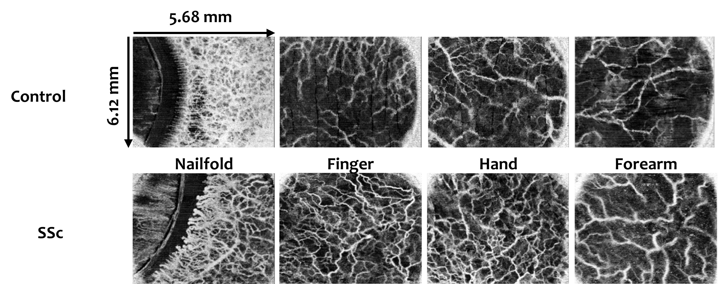
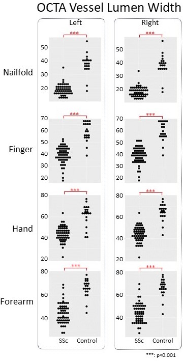
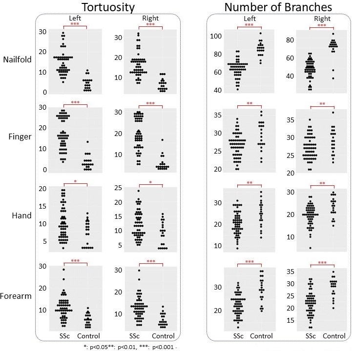
As seen in the representative images in Figure 1, OCTA was able to provide high-resolution images of cutaneous vessels in all four anatomic areas. As expected, vessel lumen width (Figure 2) and number of branches (Figure 3) were significantly lower and vascular tortuosity (Figure 3) was significantly higher in SSc patients than in healthy controls in the nailfold area. Consistent with these findings, lower vessel lumen width and number of branches, as well as higher vessel tortuosity were also observed in finger, hand, and forearm areas. The difference between SSc patients and controls was statistically significant for all comparisons. Moreover, all measurements also showed high intra- and inter-observer reliability.

Conclusion:  OCTA is a safe, non-invasive method that can assess skin vascular involvement in SSc beyond nailfold capillaries. Using this novel technology, we showed that SSc patients have prominent skin vascular changes in finger, hand, and forearm areas.   

Supporting image 1

Figure 1: Representative OCTA images of vasculature in the nailfold, finger, hand, and forearm areas of healthy control (top panel) and SSc (bottom panel) skin

Supporting image 2

Figure 2: Skin vessel lumen width in the nailfold, finger, hand, and forearm areas in SSc patients and healthy controls based on the OCTA images.

Supporting image 3

Figure 3: Skin vessel tortuosity (left panel) and number of branches (right panel) in the nailfold, finger, hand, and forearm areas in SSc patients and healthy controls based on the OCTA images.


Disclosures: P. Nikitin: None; H. Chawla: None; M. Singh: None; S. Theodore: None; S. Aglyamov: None; M. Zhang: None; B. Skaug: None; M. Mayes: AstraZeneca, 5, Atyr, 5, Boehringer-Ingelheim, 1, 5, Horizon Therapeutics, 5, Merck/MSD, 5; K. Larin: None; S. Assassi: AstraZeneca, 2, aTyr, 2, 5, BMS, 2, Boehringer-Ingelheim, 2, 5, CSL Behring, 2, Janssen, 5, Merck/MSD, 2, TeneoFour, 2.

To cite this abstract in AMA style:

Nikitin P, Chawla H, Singh M, Theodore S, Aglyamov S, Zhang M, Skaug B, Mayes M, Larin K, Assassi S. Optical Coherence Angiography: A Non-invasive, Safe Method for Assessing the Systemic Sclerosis Related Vasculopathy Beyond Nailfold Capillaries [abstract]. Arthritis Rheumatol. 2024; 76 (suppl 9). https://acrabstracts.org/abstract/optical-coherence-angiography-a-non-invasive-safe-method-for-assessing-the-systemic-sclerosis-related-vasculopathy-beyond-nailfold-capillaries/. Accessed .
  • Tweet
  • Click to email a link to a friend (Opens in new window) Email
  • Click to print (Opens in new window) Print

« Back to ACR Convergence 2024

ACR Meeting Abstracts - https://acrabstracts.org/abstract/optical-coherence-angiography-a-non-invasive-safe-method-for-assessing-the-systemic-sclerosis-related-vasculopathy-beyond-nailfold-capillaries/

Advanced Search

Your Favorites

You can save and print a list of your favorite abstracts during your browser session by clicking the “Favorite” button at the bottom of any abstract. View your favorites »

All abstracts accepted to ACR Convergence are under media embargo once the ACR has notified presenters of their abstract’s acceptance. They may be presented at other meetings or published as manuscripts after this time but should not be discussed in non-scholarly venues or outlets. The following embargo policies are strictly enforced by the ACR.

Accepted abstracts are made available to the public online in advance of the meeting and are published in a special online supplement of our scientific journal, Arthritis & Rheumatology. Information contained in those abstracts may not be released until the abstracts appear online. In an exception to the media embargo, academic institutions, private organizations, and companies with products whose value may be influenced by information contained in an abstract may issue a press release to coincide with the availability of an ACR abstract on the ACR website. However, the ACR continues to require that information that goes beyond that contained in the abstract (e.g., discussion of the abstract done as part of editorial news coverage) is under media embargo until 10:00 AM ET on November 14, 2024. Journalists with access to embargoed information cannot release articles or editorial news coverage before this time. Editorial news coverage is considered original articles/videos developed by employed journalists to report facts, commentary, and subject matter expert quotes in a narrative form using a variety of sources (e.g., research, announcements, press releases, events, etc.).

Violation of this policy may result in the abstract being withdrawn from the meeting and other measures deemed appropriate. Authors are responsible for notifying colleagues, institutions, communications firms, and all other stakeholders related to the development or promotion of the abstract about this policy. If you have questions about the ACR abstract embargo policy, please contact ACR abstracts staff at [email protected].

Wiley

  • Online Journal
  • Privacy Policy
  • Permissions Policies
  • Cookie Preferences

© Copyright 2025 American College of Rheumatology