ACR Meeting Abstracts

ACR Meeting Abstracts

  • Meetings
    • ACR Convergence 2024
    • ACR Convergence 2023
    • 2023 ACR/ARP PRSYM
    • ACR Convergence 2022
    • ACR Convergence 2021
    • ACR Convergence 2020
    • 2020 ACR/ARP PRSYM
    • 2019 ACR/ARP Annual Meeting
    • 2018-2009 Meetings
    • Download Abstracts
  • Keyword Index
  • Advanced Search
  • Your Favorites
    • Favorites
    • Login
    • View and print all favorites
    • Clear all your favorites
  • ACR Meetings

Abstract Number: 1541

MRI Digital Artery Volume Index (DAVIX) Predicts Future Digital Ulcers and Disease Progression in Systemic Sclerosis

Michael Hughes1, Stefano Di Donato2, Klodian Gjeloshi3, Giuseppina Abignano4, Fiammetta Danzo3, Giovanni Lettieri G4, Enrico De Lorenzis2, Dominic Bertham5, Philip O’Connor2, Olga Kubassova6 and Francesco Del Galdo2, 1Department of Rheumatology, Tameside and Glossop Integrated Care NHS Foundation Trust & The University of Manchester, Manchester, United Kingdom, 2Leeds Institute of Rheumatic and Musculoskeletal Medicine, Faculty of Medicine and Health and NIHR Biomedical Research Centre, University of Leeds, Leeds, United Kingdom, 3University of Campania ‘‘Luigi Vanvitelli’’, Napoli, Italy, 4Istituto Reumatologico Lucano (IReL), Azienda Ospedaliera Regionale San Carlo, Potenza, Italy, 5NIHR Leeds Biomedical Research Centre, Leeds Teaching Hospitals NHS Trust, Leeds, United Kingdom, 6Image Analysis Group, London, United Kingdom

Meeting: ACR Convergence 2022

Keywords: Imaging, Raynaud's, Systemic sclerosis, Ulcers

  • Tweet
  • Click to email a link to a friend (Opens in new window) Email
  • Click to print (Opens in new window) Print
Session Information

Date: Sunday, November 13, 2022

Title: Systemic Sclerosis and Related Disorders – Clinical Poster II

Session Type: Poster Session C

Session Time: 1:00PM-3:00PM

Background/Purpose: Fibroproliferative vasculopathy in systemic sclerosis (SSc) is caused by neointima proliferation is a key pathologic feature of SSc which results in arterial vessel narrowing (e.g., through adventitial fibrosis) and contributes to microvascular injury and damage. The aim of our study was to determine the value of non-contrast MRI-based DAVIX in predicting digital ulcers (DUs) and worsening of patient reported outcomes (PROs)/clinical manifestations in SSc.

Methods: 91 consecutive patients with Raynaud’s phenomenon were enrolled in this study.

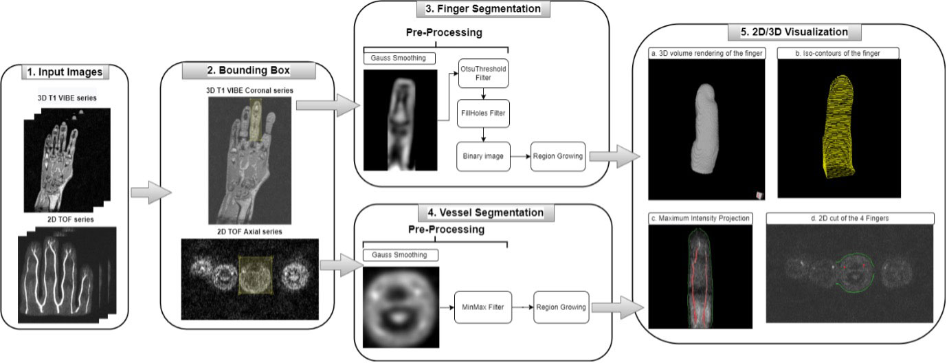
At baseline, patients underwent an MRI scan of their dominant hand. We utilised a time-of-flight MR angiography (TOF) sequence which allows the visualization of vessels by the enhancement of flowing blood and the suppression of surrounding stationary tissue in a definite volume (Figure 1). Briefly, our method uses a region growing algorithm to semi-automatically detect the arteries and the finger anatomy (Figure 1). DAVIX of the dominant hand was calculated as %mean of the 4 fingers.

Data collected included (but was not limited to): pulmonary function tests, nailfold capillaroscopy, modified Rodnan Skin Score (mRSS), history/presence of DU, and sHAQ-DI.

The distribution of data was analysed with D’Agostino-Pearson normality test. Medians were compared by Mann-Whitney-Wilcoxon test, correlation with clinical manifestations was performed using Spearman’s or Pearson test, as appropriate using Prism 7, GraphPad Software. Correlation between DAVIX and capillaroscopy was scored as a semi quantitative integer scale ranging from 0 to 4 (normal, non-specific, early, active, late).

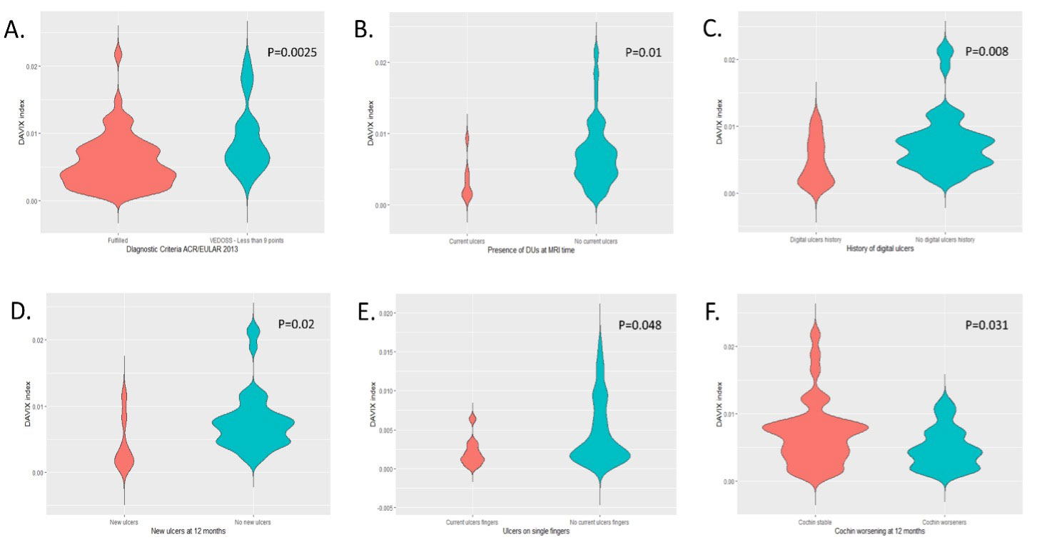
Results: The majority (86%) were females, 62 fulfilled 2013 SSc classification criteria with 5.9 (7) years median (IQR) disease duration. Twenty-nine were patients with VEDOSS (mRSS Score < 9). Complete historical and prospective (12-month) follow-up data were available for 85 patients. Patients fulfilling SSc criteria had a lower median (IQR) DAVIX compared to those with VEDOSS [0.46% (0.48) vs 0.75% (0.54), P=0.0025] (Figure 2).

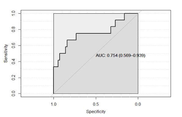
DAVIX was significantly lower in patients with DUs at baseline (P=0.01) and in those with DU history (P=0.008) (Figure 2). Further, in the group with no previous/current DU, DAVIX of patients who developed new DUs during follow-up was 0.23% vs. 0.66% (P=0.02) (Figure 2). ROC curve analysis (Figure 3) indicated that a DAVIX threshold < 0.36% conferred a 4 times higher risk of new DUs.

DAVIX correlated with disease duration (r=-0.415, P< 0.0001), baseline mRSS (r=-0.278, P=0.028), DLCO% (r=0.368, P=0.004), FVC/DLCO (r=-0.337, P=0.009), and capillaroscopy pattern (r=-0.447, P< 0.001). Additionally, DAVIX correlated with worsening HAQ-DI (r=-0.308, P=0.024), Raynaud (r=-0.270, P=0.048) and DU VAS (r=-0.291, P=0.044).

Conclusion: DAVIX is a promising surrogate outcome measure of vascular disease in SSc. The predicative value of DAVIX to predict worsening of PROs and future DUs may be useful for patient enrichment/stratification in clinical trials and offer insights into vascular disease activity in SSc disease progression.

Supporting image 1

Figure 1. Workflow of the finger and vessel segmentation. DYNAMIKA (IAG, Image Analysis Group) workflow. Left-sided images: 3D T1 VIBE images and 2D TOF images are uploaded. The target finger is identified and outlined with a bounding box. Middle images: Finger segmentation model applied to generate the volume of the finger and the vessels segmentation model is applied to generate the vessels volume. Right-sided images: 3D rendering of the segmented finger and blood vessels.

Supporting image 2

Figure 2. DAVIX in patients who fulfilled the ACR/EULAR 2013 SSc classification criteria versus patients presenting a score <9 i.e., very early diagnosis of SSc (VEDOSS) (A). DAVIX in patients with baseline digital ulcers versus patients without baseline digital ulcers (B). DAVIX in patients with history of digital ulcers versus patients who never experienced digital ulcers disease (C). DAVIX in patients without baseline digital ulcers who developed digital ulcers at 12-month follow up versus patients without baseline digital ulcers who did not develop new ulcers after 12 months (D). DAVIX of the single fingers of the dominant hands of patients presenting with at least one digital ulcer at baseline: fingers with current ulceration vs fingers without current ulceration (E). DAVIX in patients presenting a stable CHF at 12-month follow up vs patients experiencing a worsening in the CHF after 12 months (F).

Supporting image 3

Figure 3. ROC curve with area under curve for the performance of the DAVIX index in the detection of new digital ulcers at 12 months. The 0.36% cut-off shows a specificity of 84% and a sensitivity of 67% in identifying patients who went on developing new digital ulcers. Patients with a dominant hand DAVIX © index inferior to 0.36% showed a significantly higher risk of developing new ulcers at 12 months (Incident Risk Ratio=4.55, Odds Ratio=7.31, Attributable fraction in the exposed=78%, P=0.005).


Disclosures: M. Hughes, Actelion pharmaceuticals, Eli Lilly, Pfizer; S. Di Donato, None; K. Gjeloshi, None; G. Abignano, None; F. Danzo, None; G. Lettieri G, None; E. De Lorenzis, None; D. Bertham, None; P. O’Connor, None; O. Kubassova, IAG Image Analysis Group; F. Del Galdo, AbbVie/Abbott, AstraZeneca, Boehringer-Ingelheim, Mitsubishi-Tanabe, Capella biosciences, Chemomab LTD, Kymab.

To cite this abstract in AMA style:

Hughes M, Di Donato S, Gjeloshi K, Abignano G, Danzo F, Lettieri G G, De Lorenzis E, Bertham D, O’Connor P, Kubassova O, Del Galdo F. MRI Digital Artery Volume Index (DAVIX) Predicts Future Digital Ulcers and Disease Progression in Systemic Sclerosis [abstract]. Arthritis Rheumatol. 2022; 74 (suppl 9). https://acrabstracts.org/abstract/mri-digital-artery-volume-index-davix-predicts-future-digital-ulcers-and-disease-progression-in-systemic-sclerosis/. Accessed .
  • Tweet
  • Click to email a link to a friend (Opens in new window) Email
  • Click to print (Opens in new window) Print

« Back to ACR Convergence 2022

ACR Meeting Abstracts - https://acrabstracts.org/abstract/mri-digital-artery-volume-index-davix-predicts-future-digital-ulcers-and-disease-progression-in-systemic-sclerosis/

Advanced Search

Your Favorites

You can save and print a list of your favorite abstracts during your browser session by clicking the “Favorite” button at the bottom of any abstract. View your favorites »

All abstracts accepted to ACR Convergence are under media embargo once the ACR has notified presenters of their abstract’s acceptance. They may be presented at other meetings or published as manuscripts after this time but should not be discussed in non-scholarly venues or outlets. The following embargo policies are strictly enforced by the ACR.

Accepted abstracts are made available to the public online in advance of the meeting and are published in a special online supplement of our scientific journal, Arthritis & Rheumatology. Information contained in those abstracts may not be released until the abstracts appear online. In an exception to the media embargo, academic institutions, private organizations, and companies with products whose value may be influenced by information contained in an abstract may issue a press release to coincide with the availability of an ACR abstract on the ACR website. However, the ACR continues to require that information that goes beyond that contained in the abstract (e.g., discussion of the abstract done as part of editorial news coverage) is under media embargo until 10:00 AM ET on November 14, 2024. Journalists with access to embargoed information cannot release articles or editorial news coverage before this time. Editorial news coverage is considered original articles/videos developed by employed journalists to report facts, commentary, and subject matter expert quotes in a narrative form using a variety of sources (e.g., research, announcements, press releases, events, etc.).

Violation of this policy may result in the abstract being withdrawn from the meeting and other measures deemed appropriate. Authors are responsible for notifying colleagues, institutions, communications firms, and all other stakeholders related to the development or promotion of the abstract about this policy. If you have questions about the ACR abstract embargo policy, please contact ACR abstracts staff at [email protected].

Wiley

  • Online Journal
  • Privacy Policy
  • Permissions Policies
  • Cookie Preferences

© Copyright 2025 American College of Rheumatology