Session Information
Date: Monday, November 13, 2023
Title: Abstracts: Innate Immunity
Session Type: Abstract Session
Session Time: 2:00PM-3:30PM
Background/Purpose: Autoimmune photosensitivity is observed in type I Interferon (IFN) mediated diseases such as systemic and cutaneous lupus erythematosus (SLE/CLE) and dermatomyositis. Type I IFN has been described as an activator of UV-induced immune responses, but how a chronic IFN-high environment drives photosensitivity is not understood. Mitochondrial DNA has been identified as a source of IFN responses via activation of cGAS-STING in diverse cells of SLE. Here, we investigated how UV light and type I IFN exposure impact mitochondrial stress and Z-DNA formation, a left-handed dsDNA primarily localized in mitochondria which leads to type I IFN production through activation of cGAS.
Methods: Confocal microscopy of primary keratinocytes (KCs) from healthy controls and SLE patients and N/TERT immortalized KCs was performed to assess mitochondrial dynamics and cytosolic Z-DNA formation after UV exposure and IFN-α treatment. qPCR and single cell RNA sequencing was used to assess gene expression. Tissue immunofluorescence was used for protein expression of ZBP1. shRNA-mediated knockdown of ZBP1 was performed in NTERTs.
Results: After UV light exposure, NTERTs showed significantly upregulated gene expression of IFNB, IFNK, IFNL, MX1 and OASL. This upregulation was significantly inhibited by preincubation with the mitochondrially-targeted antioxidant mitoTEMPO (MT), indicating mitochondrial reactive oxygen species-dependent IFN responses. Additionally, mitochondria showed significant fragmentation after UV light that was associated with cytosolic Z-DNA accumulation. Strikingly, this accumulation was enhanced with IFN incubation leading to large Z-DNA puncta within the cytosol. Primary SLE KCs exhibit cytosolic Z-DNA at baseline and showed strong cytosolic Z-DNA accumulation after UV exposure. Cytosolic Z-DNA accumulation and UV-induced ISG expression was prevented by MT in SLE KCs. Importantly, ZBP1, the cytosolic sensor of Z-DNA, is induced by IFN-α and upregulated in nonlesional and lesional SLE and dermatomyositis skin biopsies but is not detectable in healthy control biopsies. Confocal analysis showed colocalization of Z-DNA with ZBP1 and cGAS after IFN+UVB exposure. Knockdown of ZBP1 in NTERTs attenuated ISG expression after UVB in an IFN-high environment.
Conclusion: Our data indicate that type I IFN priming, coupled with UV light exposure, results in mitochondrial stress that leads to increased mitochondrial Z-DNA formation and cytoplasmic release. Cytosolic Z-DNA interacts with ZBP1 and cGAS to activate IFN upregulation. Collectively, we describe a new pathway of mitochondrial Z-DNA sensing by ZBP1 that drives and sustains IFN responses in KCs, giving further insight into autoimmune photosensitivity.
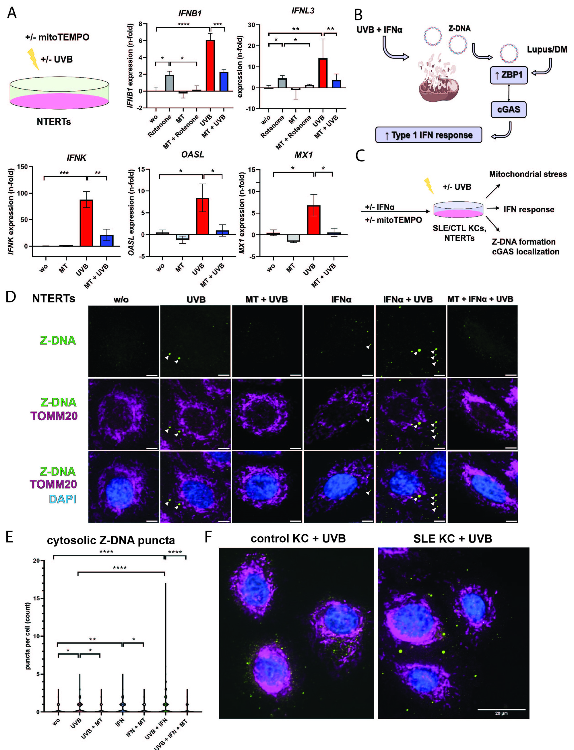
A) Expression of interferon genes in NTERTs +/- UVB exposure and +/- preincubation with mitochondrial targeted antioxidant mitoTEMPO (n=4).
B) Schematic of Z-DNA release: Upon mitochondrial damage by UVB and IFN-α, Z-DNA is released into the cytosol and bound by ZBP1 which is overexpressed in the epidermis of lupus and dermatomyositis patients. ZBP1 can interact with cGAS to sustain IFN responses in KCs.
C) Methodology to assess mitochondrial stress and Z-DNA formation in KCs.
D) Immunofluorescence (IF) staining of NTERTs identifies mitochondrial-derived cytoplasmic Z-DNA release upon UVB exposure resulting in large Z-DNA puncta after IFN-α treatment (n=4).
E) Quantification of cytoplasmic Z-DNA by automatic image analysis using CellProfiler software.
F) IF of control KCs (n=3) and SLE KCs (n=2) for Z-DNA after UVB exposure identifies large Z-DNA puncta in SLE KCs.
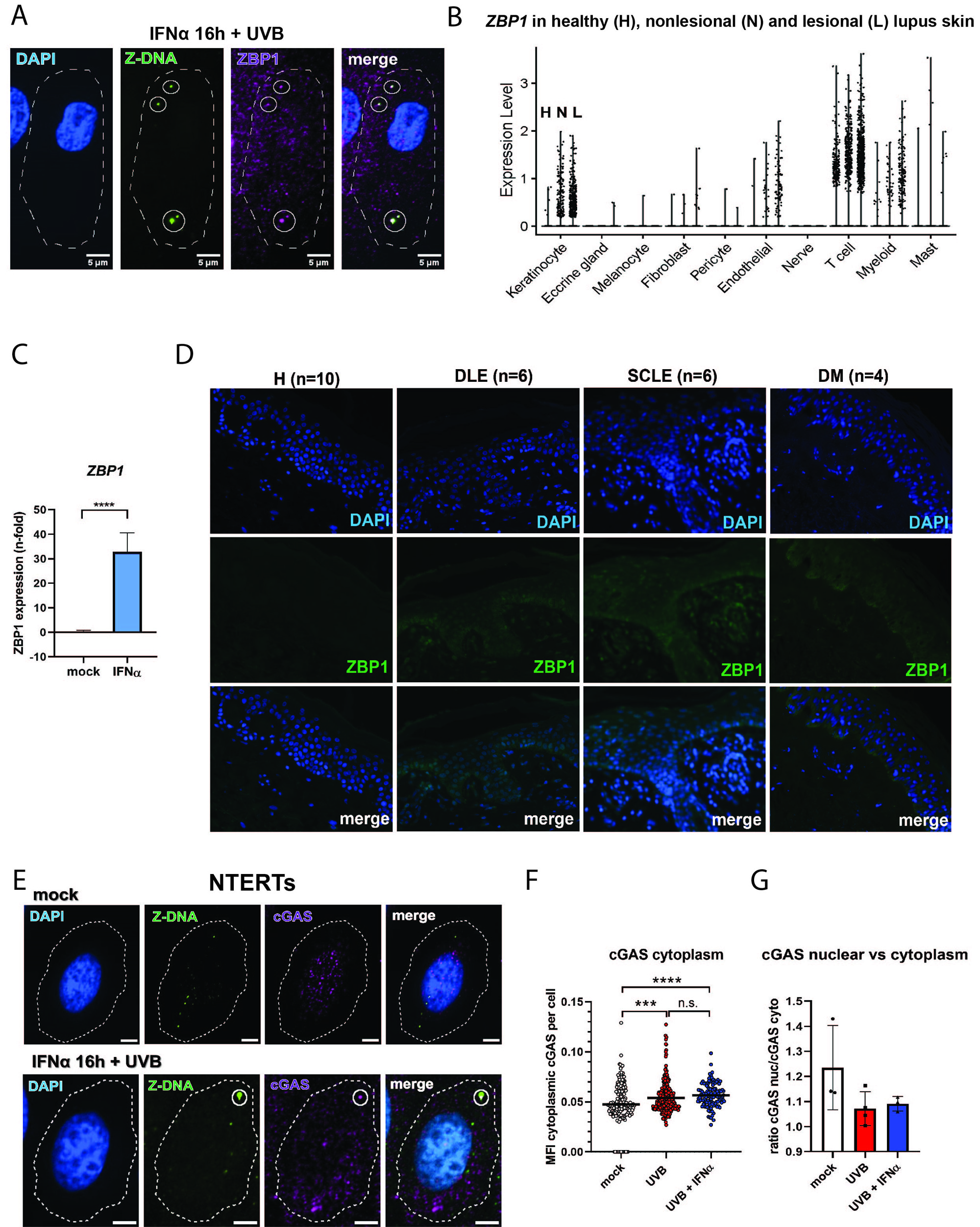
A) IF staining of Z-DNA and ZBP1 in NTERTs reveals colocalization of large cytoplasmic Z-DNA puncta with ZBP1.
B) Violin plots showing expression levels of ZBP1 in different cell types of healthy control (HC, n=14), nonlesional (n=7) and lesional (n=7) lupus skin.
C) ZBP1 is induced by IFN-α incubation in NTERTs (n=4).
D) IF staining of HC, discoid lupus (DLE), subacute cutaneous lupus (SCLE), and dermatomyositis (DM) identifies increased epidermal ZBP1 expression in autoimmune photosensitive disorders compared to HC.
E) IF staining of cGAS in NTERTs reveals colocalization of cGAS with large Z-DNA puncta after UVB exposure and IFN-α treatment (n=3).
F) Quantification of cGAS staining shows cytoplasmic shift of cGAS after UVB exposure.
G) Ratio of nuclear and cytoplasmic cGAS after UVB and IFN-α treatment.
To cite this abstract in AMA style:
Klein B, Reynolds M, Xu B, Gharaee-Kermani M, Victory A, Loftus S, O'Riordan M, Kahlenberg J. Mitochondrial Z-DNA and ZBP1 Drive Autoimmune Photosensitivity [abstract]. Arthritis Rheumatol. 2023; 75 (suppl 9). https://acrabstracts.org/abstract/mitochondrial-z-dna-and-zbp1-drive-autoimmune-photosensitivity/. Accessed .« Back to ACR Convergence 2023
ACR Meeting Abstracts - https://acrabstracts.org/abstract/mitochondrial-z-dna-and-zbp1-drive-autoimmune-photosensitivity/