Session Information
Date: Sunday, November 10, 2019
Title: T Cell Biology & Targets in Autoimmune & Inflammatory Disease Poster
Session Type: Poster Session (Sunday)
Session Time: 9:00AM-11:00AM
Background/Purpose: Anti-PD-1/PD-L1 checkpoint inhibitor therapies have produced remarkable results in stimulating immune responses against tumors. Checkpoint inhibitor therapy can also trigger a variety of autoimmune responses; however, the mechanisms underlying these immune-related adverse events (IrAEs) remain unclear. Checkpoint inhibitor-related arthritis (CIrA) occurs in ~5% of treated patients, and synovial fluid from these patients provides a unique opportunity to study the phenotypes of immune cells from the inflamed site. The extent to which the inflammatory response in CIrA resembles RA or PsA is unknown.
Methods: We conducted detailed immunophenotyping of cryopreserved synovial fluid mononuclear cells (SFMC) from CIrA following anti-PD-1/PD-L1 treatment (n=9), seropositive RA (n=5), and PsA (n=5) patients using a 39-marker mass cytometry (CyTOF) panel. CIrA patients had varied cancers, and 5/9 had mono/oligoarticular arthritis. We identified and custom-conjugated an anti-PD-1 CyTOF antibody using a clone that is not blocked by pembrolizumab or nivolumab, allowing detection of PD-1 in CIrA samples. We used FlowSOM to cluster either CD4 or CD8 T cells into 15 ‘metaclusters’ (cell populations) based on their multi-dimensional phenotypes. We used Kruskal-Wallis or Mann-Whitney tests to identify significantly altered populations (p< 0.05), which we confirmed by biaxial gating.
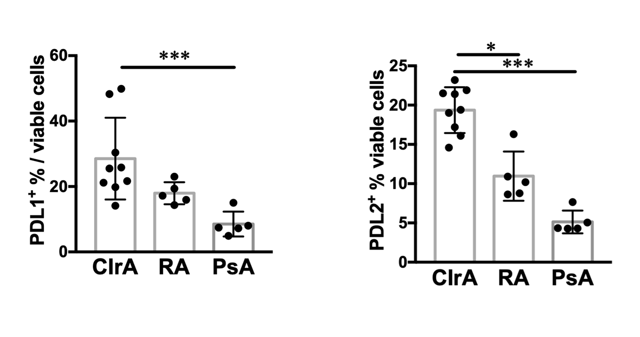
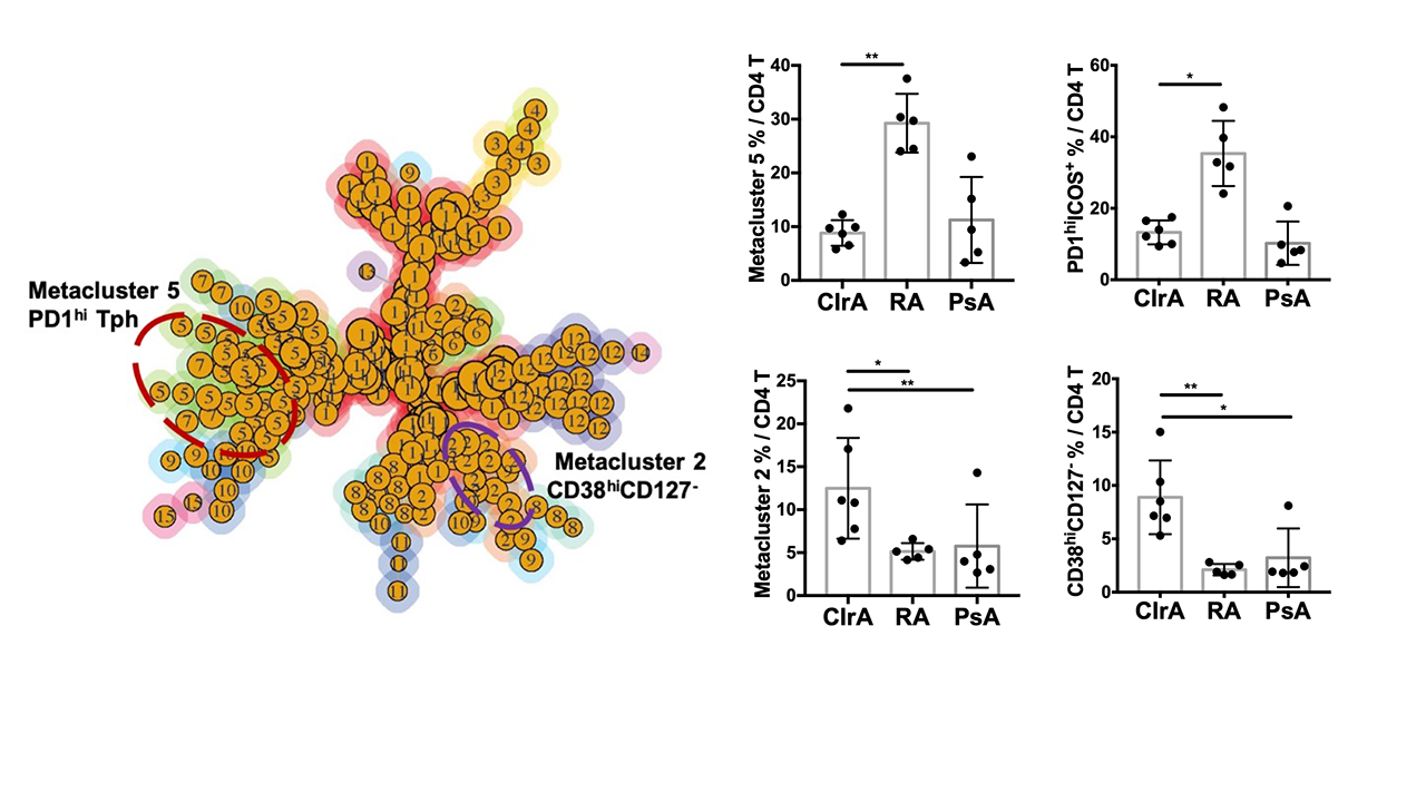
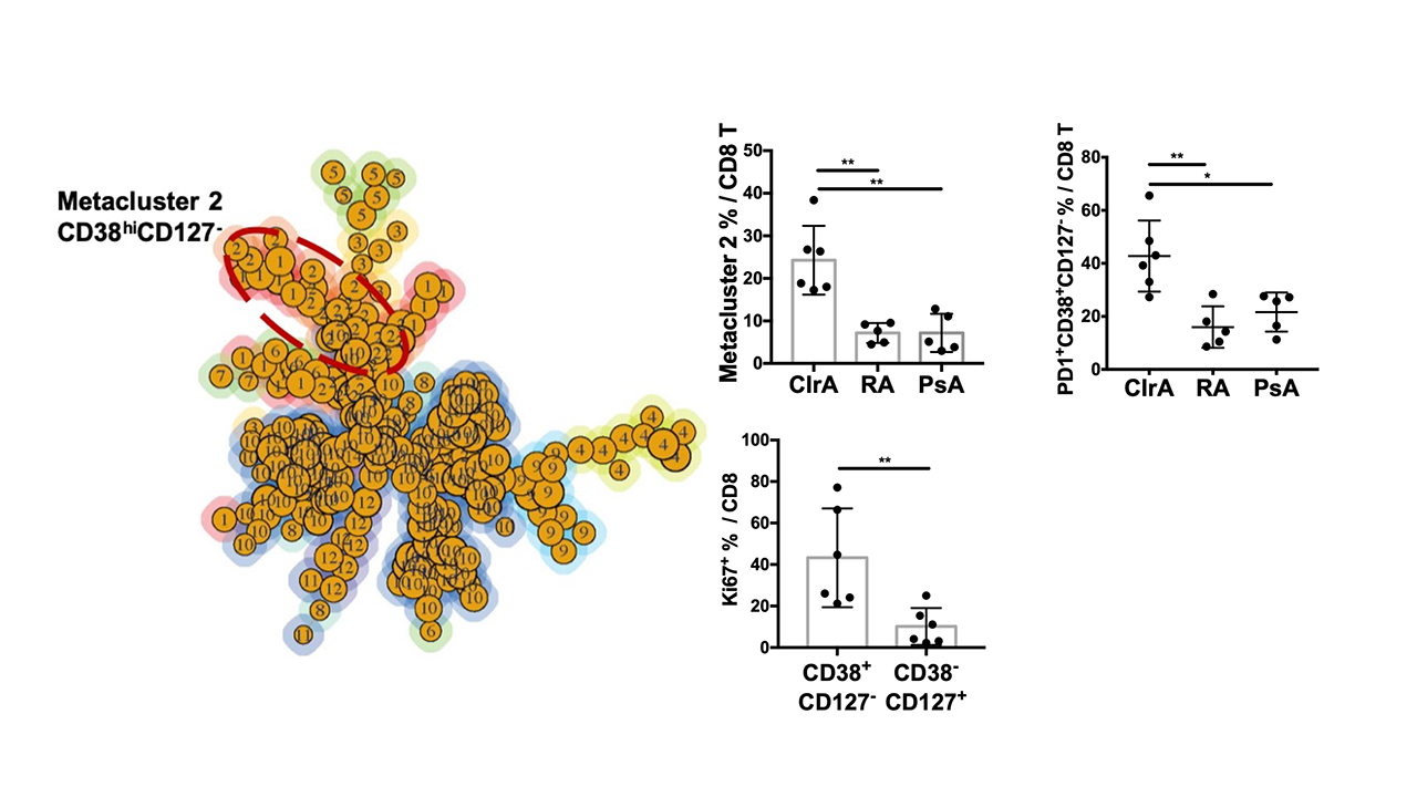
Results: T cells were the most abundant population in CIrA (50% of SFMC. 53% CD4, 40% CD8), followed by monocytes (24%) and NK cells (8%); they were comparable to those in RA and PsA. Cells in CIrA samples showed expression of PD-1 comparable to that in RA and PsA. However, expression of PD-L1 and PD-L2, both frequently expressed on monocytes, was increased in CIrA compared to RA and PsA (Fig. 1, PD-L1: 2.2-fold increased over RA/PsA, p=0.0021; PD-L2: 2.4-fold increased over RA/PsA, p< 0.0001). FlowSOM analysis of CD4 T cells highlighted distinct cell populations in CIrA and RA. RA samples contained one significantly expanded population (metacluster5, 30% of CD4s in RA, p=0.006), which contained PD-1hiICOS+ T peripheral helper (Tph) cells. Tph cells were not expanded in CIrA. In contrast, CIrA samples showed a specific expansion of a distinct population of PD-1+CD38hi CD127-cells (metacluster2, 10% of CD4s in CIrA, 2.4-fold increased over RA/PsA, p=0.0047), which we confirmed by biaxial gating (Fig. 2). Interestingly, analysis of CD8 T cells by FlowSOM revealed expansion in CIrA of a CD8 cell population with a similar phenotype to the expanded CD4 population: PD-1+CD38hiCD127-(CD8 metacluster2, 30% in CIrA, 3.4-fold increased over RA/PsA, p=0.0002). Over 40% of PD-1+CD38hi CD127-CD8+T cells in CIrA expressed Ki67, suggesting active proliferation (Fig. 3).
Conclusion: CyTOF analysis of SFMC revealed uniquely expanded CD4 and CD8 T cell populations in CIrA that are not shared with RA or PsA. Both the expanded CD4 and CD8 populations show a PD-1 + CD38 hi CD127 – phenotype. These cell phenotypes may be directly enhanced by PD-1/PD-L1 blockade and may contribute to the amplified immune response. Functional analysis of these cells may reveal key mechanisms driving CI-associated IrAEs.
To cite this abstract in AMA style:
Wang R, Chan K, Cunningham-Bussel A, Donlin L, Vitone G, Tirpack A, Benson C, Keras G, Jonsson A, Brenner M, Bass A, Rao D. Mass Cytometry Immunophenotyping of Synovial Fluid from Checkpoint Inhibitor Related Arthritis [abstract]. Arthritis Rheumatol. 2019; 71 (suppl 10). https://acrabstracts.org/abstract/mass-cytometry-immunophenotyping-of-synovial-fluid-from-checkpoint-inhibitor-related-arthritis/. Accessed .« Back to 2019 ACR/ARP Annual Meeting
ACR Meeting Abstracts - https://acrabstracts.org/abstract/mass-cytometry-immunophenotyping-of-synovial-fluid-from-checkpoint-inhibitor-related-arthritis/


