ACR Meeting Abstracts

ACR Meeting Abstracts

  • Meetings
    • ACR Convergence 2024
    • ACR Convergence 2023
    • 2023 ACR/ARP PRSYM
    • ACR Convergence 2022
    • ACR Convergence 2021
    • ACR Convergence 2020
    • 2020 ACR/ARP PRSYM
    • 2019 ACR/ARP Annual Meeting
    • 2018-2009 Meetings
    • Download Abstracts
  • Keyword Index
  • Advanced Search
  • Your Favorites
    • Favorites
    • Login
    • View and print all favorites
    • Clear all your favorites
  • ACR Meetings

Abstract Number: 2188

Lung Cysts in Primary SS: New Findings on an Allegedly Innocuous Iiagnosis

Regis Sewa Marques, Maria Fernanda Zacarin, Pamela Bellini, Raissa Dudienas D Pereira, Aloma Guinami Scabora, Zoraida Sachetto, Manoel Bertolo and ALISSON PUGLIESI, Universidade Estadual de Campinas, Campinas, Brazil

Meeting: ACR Convergence 2023

Keywords: Computed tomography (CT), Epidemiology, interstitial lung disease, pulmonary, Sjögren's syndrome

  • Tweet
  • Click to email a link to a friend (Opens in new window) Email
  • Click to print (Opens in new window) Print
Session Information

Date: Tuesday, November 14, 2023

Title: (2177–2194) Sjögren’s Syndrome – Basic & Clinical Science Poster II

Session Type: Poster Session C

Session Time: 9:00AM-11:00AM

Background/Purpose: Lung cysts are a frequent finding in patients with SS, with previous data indicating a prevalence of approximately 30% on chest CT scans. In SS, it is postulated that, among others, its genesis may be due to bronchiolar obstruction by the lymphoid tissue. Retrospective and small-sample data showed that the disease was not associated with radiological progression, suggesting an innocuous disease. However, prospective data are scarce.

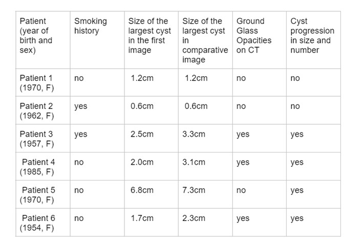
Methods: Between 2015 and 2019, patients classified as having primary SS (pSS) according to the 2002 American-European Consensus or the 2016 ACR/EULAR classification criteria and respiratory asymptomatics were systematically evaluated using high-resolution chest CT. Cysts were defined as an air space delimited by a thin (< 2 mm) layer of thin layer.Other findings were classified according to the standard descriptive guidelines. Patients with findings of pulmonary cysts on examination were subsequently evaluated with a new CT scan within at least 1 year, using the same CT machine, and analyzed by the same thoracic radiologist. The cysts were characterized comparatively by their largest diameters and numerically. Patients who increased in size by at least 0.5 cm and the total number of cysts were considered to have radiological progression.

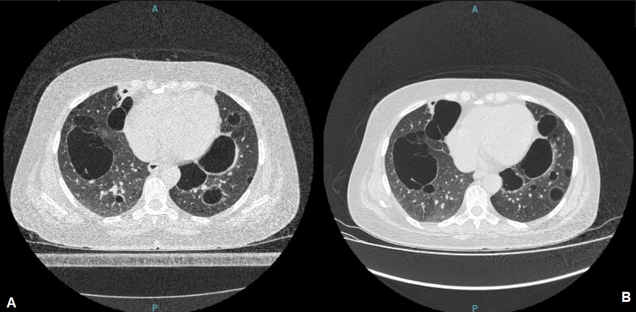
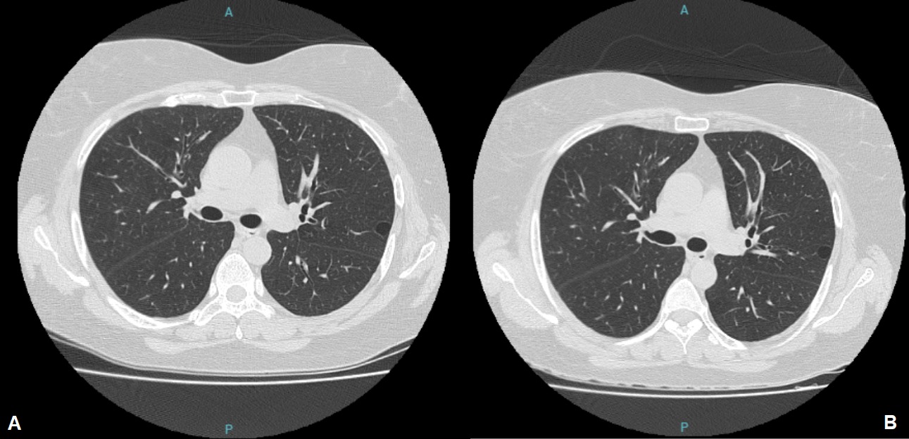
Results: Thirty-five patients with pSS without respiratory symptoms were evaluated using high-resolution chest CT. Of these, 11 (31.4%) had at least one lung cyst, and in six, a prospective evaluation was performed (the remaining patients were not evaluated because of difficulties caused by the Covid-19 pandemic). Among them, four patients showed an increase in both the size and quantity of the cysts, especially those with cysts larger than 2 cm in diameter and with ground-glass opacities on CT. Two patients who had fewer than five cysts each, all smaller than 1.3 cm, had no radiological evolution. In figure 1, we summarize the main radiological characteristics of the evaluated patients, and in figures 2 and 3, we illustrate two patients with lung cysts but with different evolutions.

Conclusion: We found a high prevalence (31.4%) of lung cysts in asymptomatic respiratory patients with pSS who underwent chest CT scans, with most showing radiological progression. In our small sample, patients with cysts > 2 cm or those with ground-glass opacity showed radiological progression. Our findings do not support the hypothesis that lung cysts in patients with pSS do not progress, and point to the need for further larger studies to better evaluate this common lung manifestation of pSS.

Supporting image 1

Table 1: Main radiological characteristics of prospectively evaluated patients with pulmonary cysts

Supporting image 2

Figure 1 – Thoracic imaging of a 52-year-old woman with cystic lung disease. (A) CT scan done in 2015 showing multiple lung cysts. (B) Progression of cysts in number and size on CT scan taken in 2018

Supporting image 3

Figure 2 – Thoracic imaging of a 52-year-old woman with cystic lung disease. (A) CT scan done in 2018 with a lung cyst in left lung lobe. (B) New CT scan done in 2019 with no progression of the cyst.


Disclosures: R. Sewa Marques: None; M. Zacarin: None; P. Bellini: None; R. Dudienas D Pereira: None; A. Guinami Scabora: None; Z. Sachetto: None; M. Bertolo: None; A. PUGLIESI: None.

To cite this abstract in AMA style:

Sewa Marques R, Zacarin M, Bellini P, Dudienas D Pereira R, Guinami Scabora A, Sachetto Z, Bertolo M, PUGLIESI A. Lung Cysts in Primary SS: New Findings on an Allegedly Innocuous Iiagnosis [abstract]. Arthritis Rheumatol. 2023; 75 (suppl 9). https://acrabstracts.org/abstract/lung-cysts-in-primary-ss-new-findings-on-an-allegedly-innocuous-iiagnosis/. Accessed .
  • Tweet
  • Click to email a link to a friend (Opens in new window) Email
  • Click to print (Opens in new window) Print

« Back to ACR Convergence 2023

ACR Meeting Abstracts - https://acrabstracts.org/abstract/lung-cysts-in-primary-ss-new-findings-on-an-allegedly-innocuous-iiagnosis/

Advanced Search

Your Favorites

You can save and print a list of your favorite abstracts during your browser session by clicking the “Favorite” button at the bottom of any abstract. View your favorites »

All abstracts accepted to ACR Convergence are under media embargo once the ACR has notified presenters of their abstract’s acceptance. They may be presented at other meetings or published as manuscripts after this time but should not be discussed in non-scholarly venues or outlets. The following embargo policies are strictly enforced by the ACR.

Accepted abstracts are made available to the public online in advance of the meeting and are published in a special online supplement of our scientific journal, Arthritis & Rheumatology. Information contained in those abstracts may not be released until the abstracts appear online. In an exception to the media embargo, academic institutions, private organizations, and companies with products whose value may be influenced by information contained in an abstract may issue a press release to coincide with the availability of an ACR abstract on the ACR website. However, the ACR continues to require that information that goes beyond that contained in the abstract (e.g., discussion of the abstract done as part of editorial news coverage) is under media embargo until 10:00 AM ET on November 14, 2024. Journalists with access to embargoed information cannot release articles or editorial news coverage before this time. Editorial news coverage is considered original articles/videos developed by employed journalists to report facts, commentary, and subject matter expert quotes in a narrative form using a variety of sources (e.g., research, announcements, press releases, events, etc.).

Violation of this policy may result in the abstract being withdrawn from the meeting and other measures deemed appropriate. Authors are responsible for notifying colleagues, institutions, communications firms, and all other stakeholders related to the development or promotion of the abstract about this policy. If you have questions about the ACR abstract embargo policy, please contact ACR abstracts staff at [email protected].

Wiley

  • Online Journal
  • Privacy Policy
  • Permissions Policies
  • Cookie Preferences

© Copyright 2025 American College of Rheumatology