ACR Meeting Abstracts

ACR Meeting Abstracts

  • Meetings
    • ACR Convergence 2024
    • ACR Convergence 2023
    • 2023 ACR/ARP PRSYM
    • ACR Convergence 2022
    • ACR Convergence 2021
    • ACR Convergence 2020
    • 2020 ACR/ARP PRSYM
    • 2019 ACR/ARP Annual Meeting
    • 2018-2009 Meetings
    • Download Abstracts
  • Keyword Index
  • Advanced Search
  • Your Favorites
    • Favorites
    • Login
    • View and print all favorites
    • Clear all your favorites
  • ACR Meetings

Abstract Number: 0812

Length of Synovial Fluid Monosodium Urate Crystals According to Sonographic Articular Deposits: Advancing in the Crystallization Process

Elena Sansano1, Mª Carmen López-González2, Cristina Rodríguez-Alvear3, Irene Calabuig-Sais3, Agustín Martínez-Sanchís3, Eliseo Pascual3 and Mariano Andrés3, 1Miguel Hernandez University, Alicante, Spain, 2General University Hospital Dr. Balmis, Alacante, Spain, 3Dr Balmis Alicante General University Hospital-ISABIAL, Alicante, Spain

Meeting: ACR Convergence 2023

Keywords: Crystal-induced arthritis, gout, Imaging

  • Tweet
  • Click to email a link to a friend (Opens in new window) Email
  • Click to print (Opens in new window) Print
Session Information

Date: Sunday, November 12, 2023

Title: Abstracts: Metabolic & Crystal Arthropathies – Basic & Clinical Science

Session Type: Abstract Session

Session Time: 4:00PM-5:30PM

Background/Purpose: We described bands of fibers having deposited orderly arrayed monosodium urate (MSU) crystals suggesting the need for protein templates to start crystallization [PMID 9709185]. However, tophi sections demonstrate the presence of longer crystals, highly arranged in a fan display, likely formed after other crystals [PMID 26369610]. Hence, a double mechanism of crystallization may exist in the joint. Here, we aimed to assess whether synovial fluid MSU crystals found in joints with organized sonographic deposits (likely, many crystals deriving from them) are longer than when deposits are absent or not visible.

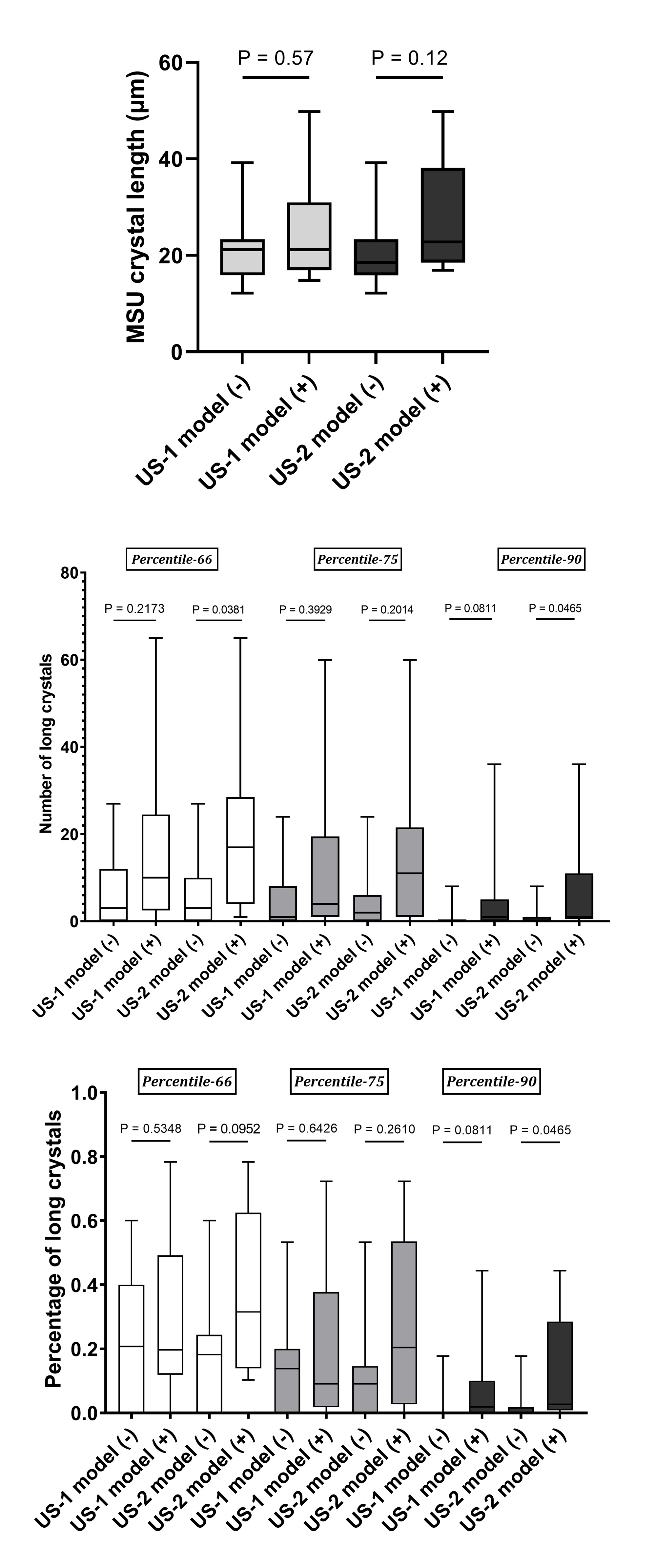
Methods: We recruited patients with crystal-proven gout during a flare. Ultrasound of the target joint was performed to detect elementary gout lesions [OMERACT 2021]: double-contour (DC) sign, tophi, and aggregates. Two sonographic models were assessed: US-1 model, grade 2-3 of any deposit (DC sign, tophi, or aggregates), and US-2 model, limited to grade 2-3 DC sign or tophi. Later, synovial fluid was aspirated. The length of MSU crystals was measured by a blinded observer using a calibrated polarized light microscope. The distribution of MSU crystal length (in µm) and the presence of long crystals (defined according to percentiles 66, 75, or 90) were tested with each sonographic model using Mann-Whitney U’s test. Quantitative variables are shown as median (p25-75).

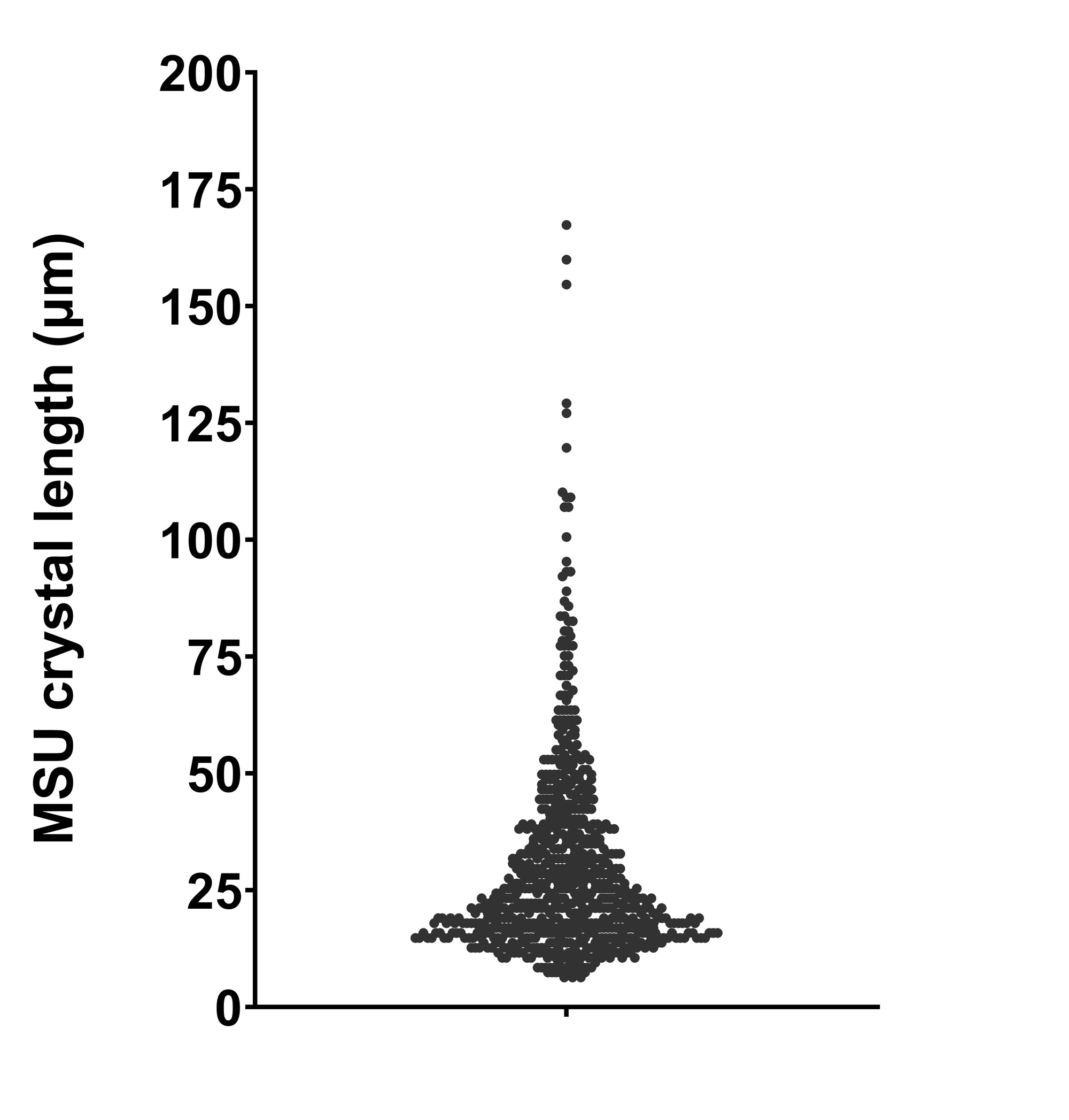
Results: We analyzed 742 crystals from 20 joints in 17 patients (three with an oligo-polyarticular flare), median aged 62.5 years (55-73), and 30% with subcutaneous tophi. The 2-year median serum urate was 7.5mg/dL (7.1-8.2), with current urate-lowering therapy in 35% of patients. The median length of MSU crystals was 21.2µm (95%CI 17.7-26.5), where crystals mostly gathered [Figure 1]. Applying the US-1 model (fulfilled by 13 joints, 65%), no difference in crystal lengths was noted (21.2µm in those with deposits vs. 21.2µm in those without deposits; p=0.588) [Figure 2, top], with a similar distribution of long crystals (P66, P75 or P90) [Figure 2, middle and bottom]. However, those with deposits according to US-2 model (n=9, 45%) showed a numerically greater crystal length (22.8µm vs. 18.5µm; p=0.112) [Figure 2, top] and more presence of long crystals [Figure 2, middle and bottom] compared to patients without deposits. Crystal lengths and the presence of long crystals showed no association with gout characteristics or serum urate levels.

Conclusion: Our synovial fluid analysis from different individuals with gout suggests two different mechanisms of MSU crystallization, one of reduced length shared by most crystals and the other with longer crystals better identified by the presence of sonographic deposits (but not aggregates), which resembles what is seen in tophi pathological sections. This exploratory finding adds evidence to the hypothesis that a secondary formation using as templates previously formed crystals occurs in the process of MSU crystallization.

Supporting image 1

Distribution of monosodium urate crystals length in the whole sample.

Supporting image 2

Comparisons between ultrasound models in crystal length (top), number of long crystals – according to percentiles 66, 75, or 90 – (middle), and percentage of long crystals – according to percentiles 66, 75, or 90 (bottom).


Disclosures: E. Sansano: None; M. López-González: None; C. Rodríguez-Alvear: None; I. Calabuig-Sais: None; A. Martínez-Sanchís: None; E. Pascual: None; M. Andrés: None.

To cite this abstract in AMA style:

Sansano E, López-González M, Rodríguez-Alvear C, Calabuig-Sais I, Martínez-Sanchís A, Pascual E, Andrés M. Length of Synovial Fluid Monosodium Urate Crystals According to Sonographic Articular Deposits: Advancing in the Crystallization Process [abstract]. Arthritis Rheumatol. 2023; 75 (suppl 9). https://acrabstracts.org/abstract/length-of-synovial-fluid-monosodium-urate-crystals-according-to-sonographic-articular-deposits-advancing-in-the-crystallization-process/. Accessed .
  • Tweet
  • Click to email a link to a friend (Opens in new window) Email
  • Click to print (Opens in new window) Print

« Back to ACR Convergence 2023

ACR Meeting Abstracts - https://acrabstracts.org/abstract/length-of-synovial-fluid-monosodium-urate-crystals-according-to-sonographic-articular-deposits-advancing-in-the-crystallization-process/

Advanced Search

Your Favorites

You can save and print a list of your favorite abstracts during your browser session by clicking the “Favorite” button at the bottom of any abstract. View your favorites »

All abstracts accepted to ACR Convergence are under media embargo once the ACR has notified presenters of their abstract’s acceptance. They may be presented at other meetings or published as manuscripts after this time but should not be discussed in non-scholarly venues or outlets. The following embargo policies are strictly enforced by the ACR.

Accepted abstracts are made available to the public online in advance of the meeting and are published in a special online supplement of our scientific journal, Arthritis & Rheumatology. Information contained in those abstracts may not be released until the abstracts appear online. In an exception to the media embargo, academic institutions, private organizations, and companies with products whose value may be influenced by information contained in an abstract may issue a press release to coincide with the availability of an ACR abstract on the ACR website. However, the ACR continues to require that information that goes beyond that contained in the abstract (e.g., discussion of the abstract done as part of editorial news coverage) is under media embargo until 10:00 AM ET on November 14, 2024. Journalists with access to embargoed information cannot release articles or editorial news coverage before this time. Editorial news coverage is considered original articles/videos developed by employed journalists to report facts, commentary, and subject matter expert quotes in a narrative form using a variety of sources (e.g., research, announcements, press releases, events, etc.).

Violation of this policy may result in the abstract being withdrawn from the meeting and other measures deemed appropriate. Authors are responsible for notifying colleagues, institutions, communications firms, and all other stakeholders related to the development or promotion of the abstract about this policy. If you have questions about the ACR abstract embargo policy, please contact ACR abstracts staff at [email protected].

Wiley

  • Online Journal
  • Privacy Policy
  • Permissions Policies
  • Cookie Preferences

© Copyright 2025 American College of Rheumatology