ACR Meeting Abstracts

ACR Meeting Abstracts

  • Meetings
    • ACR Convergence 2024
    • ACR Convergence 2023
    • 2023 ACR/ARP PRSYM
    • ACR Convergence 2022
    • ACR Convergence 2021
    • ACR Convergence 2020
    • 2020 ACR/ARP PRSYM
    • 2019 ACR/ARP Annual Meeting
    • 2018-2009 Meetings
    • Download Abstracts
  • Keyword Index
  • Advanced Search
  • Your Favorites
    • Favorites
    • Login
    • View and print all favorites
    • Clear all your favorites
  • ACR Meetings

Abstract Number: 2550

Infiltrating and Resident Macrophages in Lupus Nephritis: Scar Associated Macrophage Phagocytic Dysfunction and Fibroblast Activation

Chirag Raparia1, Paul Hoover2, Arnon Arazi3, Nir Hacohen4 and Anne Davidson5, 1Donald and Barbara Zucker School of Medicine At Hofstra/Northwell, Shoreham, NY, 2Brigham and Women's Hospital, SWAMPSCOTT, MA, 3Feinstein Institutes for Medical Research, Melrose, MA, 4Broad Institute of MIT and Harvard, Boston, MA, 5Feinstein Institutes for Medical Research, Manhasset, NY

Meeting: ACR Convergence 2024

Keywords: autoimmune diseases, Fibroblasts, Other, Lupus nephritis, macrophages, Mouse Models, Lupus

  • Tweet
  • Click to email a link to a friend (Opens in new window) Email
  • Click to print (Opens in new window) Print
Session Information

Date: Monday, November 18, 2024

Title: Abstracts: SLE – Animal Models

Session Type: Abstract Session

Session Time: 1:00PM-2:30PM

Background/Purpose: Lupus nephritis (LN) is driven by a heterogeneous population of renal macrophages. We have previously reported that the renal macrophage subpopulations are similar in mouse and human LN. In both species, disease-associated sub-populations of infiltrating (classical 2) and resident macrophages correlate with higher LN activity and chronicity scores respectively and express genes associated with Scar Associated Macrophages (SAMs) including CTSB, SPP1, FABP5, TREM2, and CD63. However, the function of these cells in LN kidneys are still unknown. Here, we elucidate the phagocytic capacity and effects of these disease-associated macrophage subsets on fibroblast cells in-vitro.

Methods: We used flow cytometry to identify macrophage subsets in nephritic kidneys of two commonly employed lupus mouse models, NZB/W and SLE1.Yaa. Phagocytic capacity was measured by flow cytometry after cell exposure to immune complexes, apoptotic cells and Latex beads ex vivo. Because infiltrating SAMs constitute only a small population of renal cells, we developed an in-vitro culture system by polarizing SLE1.Yaa bone marrow-derived macrophages (BMDMs) with GM-CSF and IL-17A, resulting in induced SAMs (iSAMs) and confirmed their phenotype with flow cytometry and RNA sequencing. The ability of iSAM-derived supernatants to promote wound repair was assessed through fibroblast migration and fibroblast density in a scratch wound assay.  Similar experiments were performed using resident macrophages isolated from either pre-nephritic or nephritic LN kidneys.

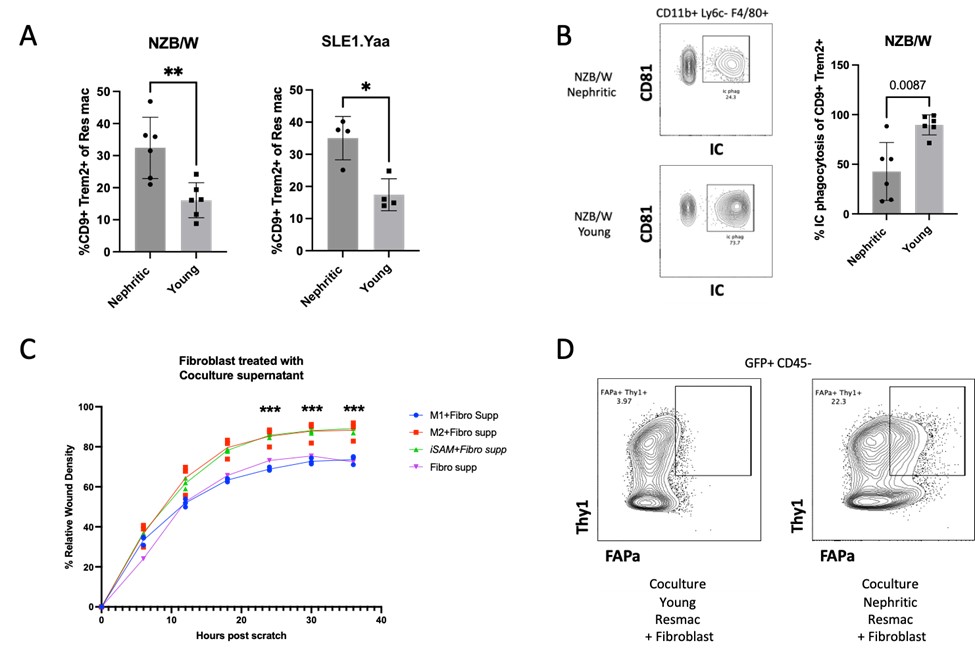
Results: Nephritic mice had a higher frequency and number of both infiltrating and resident macrophage (Fig 1A) CD9+ Trem2+ SAMs compared to non-nephritic controls. Nephritic resident macrophages had a profound defect in phagocytosis of immune complexes (Fig 1B) and apoptotic cells but not of latex beads. Phagocytosis was not defective in renal classical 1 or classical 2 macrophages from nephritic mice. The in-vitro-derived GM-CSF and IL-17A and M2 polarized BMDM expressed significantly more CD9 and Trem2 compared to M0 and M1 macrophages. When the scratch wound was exposed to the iSAM-fibroblast co-culture supernatant, fibroblast migration, and wound density were increased in both GM-CSF and IL-17A and M2 polarized BMDM compared to M0 and M1 (Fig 1C).  Nephritic resident macrophages enhanced fibroblast density within the wound area compared to pre-nephritic resident macrophages and induced an activated FAP+ fibroblast phenotype (Fig 1D).

Conclusion: Infiltrating and resident macrophages that correlate with lupus nephritis disease severity include SAMs. Resident macrophages but not infiltrating macrophages from nephritic mice display a receptor-mediated phagocytosis defect. When SAMs are derived in-vitro with IL-17a and GM-CSF their supernatant enhances fibroblast migration and density within the wound. Similarly, resident macrophages from nephritic mice display a functional pro-fibrotic phenotype compared with those from pre-nephritic mice. These studies suggest that therapeutic targeting of SAMs may attenuate fibrosis in LN.  

Supporting image 1

Figure 1 In-vivo and in-vitro characterization of Scar Associated Macrophages (SAMs) in Lupus Nephritis. A. CD9+ Trem2+ Resident Macrophages in Nephritic and Young NZB/W and SLE1.Yaa mice. B. Ex-vivo Immune Complex phagocytosis by Resident Macrophages in Nephritic and young NZB/W mice. C. Relative Wound Density of fibroblast treated with SAM, M1, and M2 fibroblast cocultured supernatant. D. FAPa+ Thy1+ fibroblast from coculture with young and nephritic Resident macrophages


Disclosures: C. Raparia: None; P. Hoover: None; A. Arazi: None; N. Hacohen: None; A. Davidson: EMD Serono, 5.

To cite this abstract in AMA style:

Raparia C, Hoover P, Arazi A, Hacohen N, Davidson A. Infiltrating and Resident Macrophages in Lupus Nephritis: Scar Associated Macrophage Phagocytic Dysfunction and Fibroblast Activation [abstract]. Arthritis Rheumatol. 2024; 76 (suppl 9). https://acrabstracts.org/abstract/infiltrating-and-resident-macrophages-in-lupus-nephritis-scar-associated-macrophage-phagocytic-dysfunction-and-fibroblast-activation/. Accessed .
  • Tweet
  • Click to email a link to a friend (Opens in new window) Email
  • Click to print (Opens in new window) Print

« Back to ACR Convergence 2024

ACR Meeting Abstracts - https://acrabstracts.org/abstract/infiltrating-and-resident-macrophages-in-lupus-nephritis-scar-associated-macrophage-phagocytic-dysfunction-and-fibroblast-activation/

Advanced Search

Your Favorites

You can save and print a list of your favorite abstracts during your browser session by clicking the “Favorite” button at the bottom of any abstract. View your favorites »

All abstracts accepted to ACR Convergence are under media embargo once the ACR has notified presenters of their abstract’s acceptance. They may be presented at other meetings or published as manuscripts after this time but should not be discussed in non-scholarly venues or outlets. The following embargo policies are strictly enforced by the ACR.

Accepted abstracts are made available to the public online in advance of the meeting and are published in a special online supplement of our scientific journal, Arthritis & Rheumatology. Information contained in those abstracts may not be released until the abstracts appear online. In an exception to the media embargo, academic institutions, private organizations, and companies with products whose value may be influenced by information contained in an abstract may issue a press release to coincide with the availability of an ACR abstract on the ACR website. However, the ACR continues to require that information that goes beyond that contained in the abstract (e.g., discussion of the abstract done as part of editorial news coverage) is under media embargo until 10:00 AM ET on November 14, 2024. Journalists with access to embargoed information cannot release articles or editorial news coverage before this time. Editorial news coverage is considered original articles/videos developed by employed journalists to report facts, commentary, and subject matter expert quotes in a narrative form using a variety of sources (e.g., research, announcements, press releases, events, etc.).

Violation of this policy may result in the abstract being withdrawn from the meeting and other measures deemed appropriate. Authors are responsible for notifying colleagues, institutions, communications firms, and all other stakeholders related to the development or promotion of the abstract about this policy. If you have questions about the ACR abstract embargo policy, please contact ACR abstracts staff at [email protected].

Wiley

  • Online Journal
  • Privacy Policy
  • Permissions Policies
  • Cookie Preferences

© Copyright 2025 American College of Rheumatology