Session Information
Session Type: ACR Concurrent Abstract Session
Session Time: 2:30PM-4:00PM
Background/Purpose: Previously we found a strong enrichment of highly expanded
T-cell clones in rheumatoid arthritis (RA) synovial tissue of inflamed joints. To
gain more insight into the potential role of these expanded clones in the
inflammatory polyarthritis in this disease, we studied the distribution of these
T-cell clones: Are they present only locally in the inflamed synovium? If not,
to what extent are they also present in synovial biopsies taken from a different
location in the same joint, or from a different joint?
Methods: In
seven RA-patients we simultaneously obtained synovial tissue (ST) biopsies from
two inflamed joints (N=7; ankle or knee) via mini-arthroscopy together with paired
peripheral blood (PB; N=7) and synovial fluid samples (SF; N=5). ST biopsies
were obtained from ankle (N=1) or from the infrapatellar region of the knee
(N=6). In 6 patients we took
additional independent suprapatellar (SP) biopsies within the same knee joint. Samples
were processed for RNA-based next generation sequencing (NGS). T-cell clones
were identified by their unique T-cell receptor β-chain sequence, the degree
of expansion being expressed as a percentage of the total number of NGS reads. (Paired)
student T-tests or Wilcoxon (matched-pair) signed rank test were performed
where applicable.
Results: We
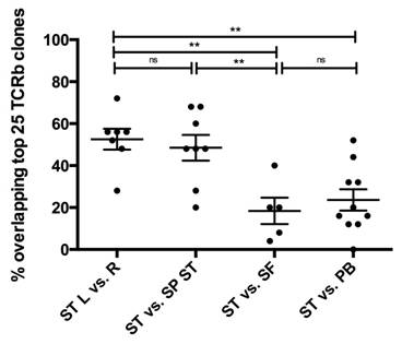
identified 904,631 clones in 6,955,333 TCR-sequences. Of the top-25 most
expanded clones in ST only a limited number were retrieved as top-25 clone in
SF (18%; SEM 6.3) and in PB (24%,
SEM 5.1). In contrast, considerable overlap was seen if we analyzed cellular
infiltrate in synovial tissue biopsies from different regions of the same joint:
49% (SEM 6.2) of the top-25 ST clones were also retrieved as a top-25 clone in
the suprapatellar biopsy of the same joint (p<0.005).
When comparing different joints in the same patient, we noted that 52% (SEM 5.0) of the top-25 clones from the 1st
joint were also amongst the top-25 clones in the 2nd joint (see
figure 1).
Conclusion: Synovial
fluid T-cells do not fully reflect the repertoire of dominant clones in the
synovial tissue. Synovial tissue biopsies from different locations within the
same joint or taken from different joints show substantial overlap of the most
dominant T-cell clones in patients with active RA. Given the evidence pointing
to T-cell involvement in the pathogenesis of RA this suggests that in
individual patients a limited number of T-cell clones dominate the adaptive
immune response. Detailed cellular characterization
of these clones seems warranted, and may help to devise more selective
strategies for therapeutic targeting.
Figure 1 |
percentage of overlapping top 25 TCRb clones. ST= synovial tissue; L= left; R=
right; SP; suprapatellar region; SF= synovial fluid; PB= peripheral blood. ** =
p<0.005; ns= not significant.
To cite this abstract in AMA style:
Musters A, Klarenbeek PL, Doorenspleet ME, Esveldt REE, van Schaik BDC, Tas SW, van Kampen AHC, Baas F, de Vries N. In Rheumatoid Arthritis Only a Few Expanded T-Cell Clones Dominate in Joint Inflammation: A Study in Seven RA Patients Undergoing Paired Synovial Tissue Biopsies in Multiple Joints [abstract]. Arthritis Rheumatol. 2015; 67 (suppl 10). https://acrabstracts.org/abstract/in-rheumatoid-arthritis-only-a-few-expanded-t-cell-clones-dominate-in-joint-inflammation-a-study-in-seven-ra-patients-undergoing-paired-synovial-tissue-biopsies-in-multiple-joints/. Accessed .« Back to 2015 ACR/ARHP Annual Meeting
ACR Meeting Abstracts - https://acrabstracts.org/abstract/in-rheumatoid-arthritis-only-a-few-expanded-t-cell-clones-dominate-in-joint-inflammation-a-study-in-seven-ra-patients-undergoing-paired-synovial-tissue-biopsies-in-multiple-joints/

