ACR Meeting Abstracts

ACR Meeting Abstracts

  • Meetings
    • ACR Convergence 2024
    • ACR Convergence 2023
    • 2023 ACR/ARP PRSYM
    • ACR Convergence 2022
    • ACR Convergence 2021
    • ACR Convergence 2020
    • 2020 ACR/ARP PRSYM
    • 2019 ACR/ARP Annual Meeting
    • 2018-2009 Meetings
    • Download Abstracts
  • Keyword Index
  • Advanced Search
  • Your Favorites
    • Favorites
    • Login
    • View and print all favorites
    • Clear all your favorites
  • ACR Meetings

Abstract Number: 1625

Identifying Potential Neuroimaging Biomarkers of Neuropsychiatric Lupus in Children Using Deep Learning

santiago arciniegas1, Matthias W. Wagner2, Logi Vidarsson2, Birgit Ertl-Wagner2, Tala El Tal3, Asha Jeyanathan2, Lawrence Ng2, Linda Hiraki2, Deborah Levy2, George M. Ibrahim2 and Andrea Knight2, 1University of Toronto, Toronto, ON, Canada, 2The Hospital for Sick Children, Toronto, ON, Canada, 3Children's Hospital of Eastern Ontario (CHEO), Ottawa, ON, Canada

Meeting: ACR Convergence 2023

Keywords: Biomarkers, Neuroimaging, neuropsychiatric disorders, Pediatric rheumatology, Systemic lupus erythematosus (SLE)

  • Tweet
  • Click to email a link to a friend (Opens in new window) Email
  • Click to print (Opens in new window) Print
Session Information

Date: Monday, November 13, 2023

Title: Abstracts: Pediatric Rheumatology – Clinical II: Connective Tissue Disease

Session Type: Abstract Session

Session Time: 2:00PM-3:30PM

Background/Purpose: Systemic lupus erythematosus (SLE) is a chronic, multisystem, autoimmune disease. Childhood-onset SLE (cSLE) is characterised by a higher frequency of nervous system involvement, known as neuropsychiatric SLE (NPSLE). Biomarkers for NPSLE are lacking, posing challenges for clinical diagnosis and management. The existence of neuropsychiatric manifestations in patients without a definite NPSLE diagnosis, as well as normal conventional magnetic resonance imaging (MRI) findings in patients with NPSLE, allude to suboptimal diagnostic protocols. To identify potential biomarkers of NPSLE, we aimed to develop a deep learning model capable of identifying structural brain image features of patients with cSLE.

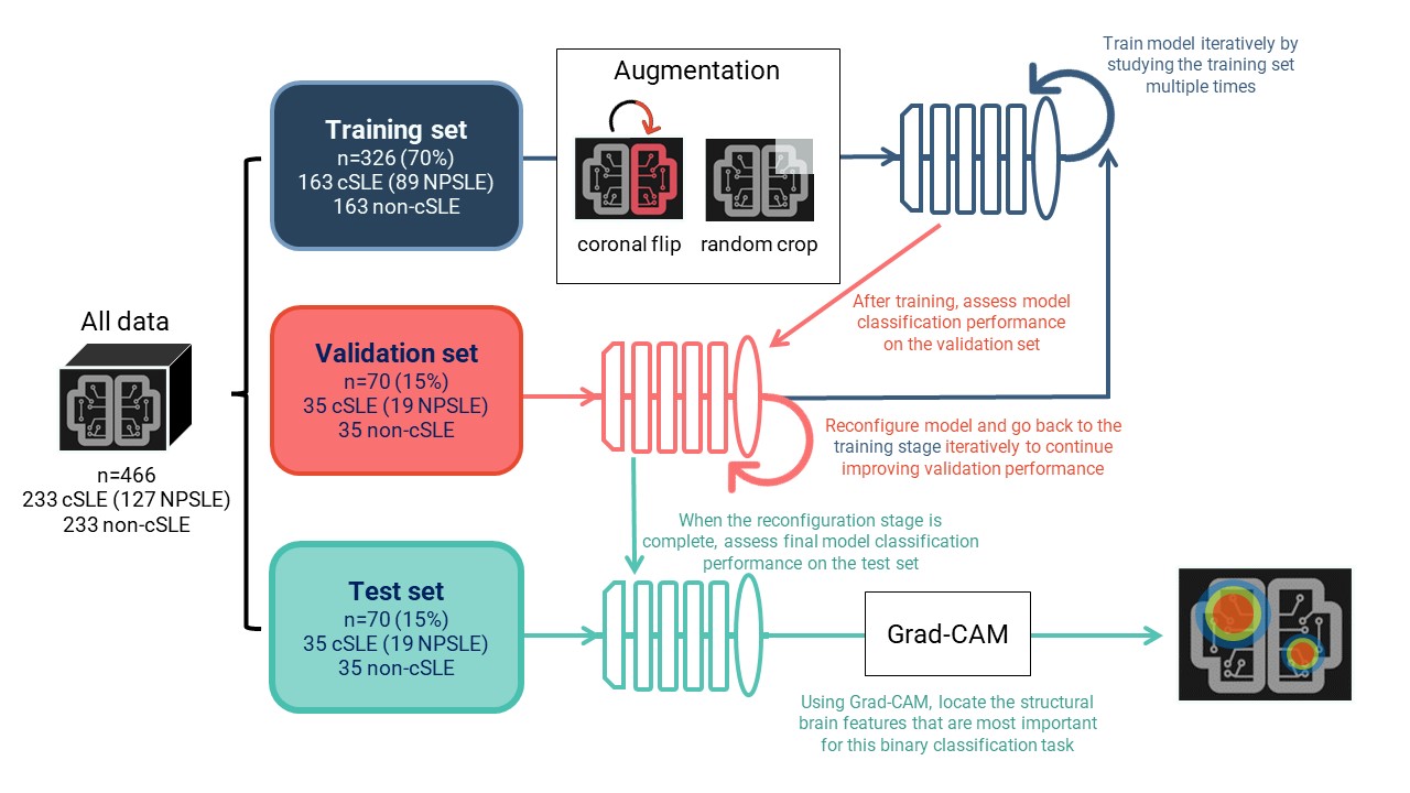
Methods: This retrospective study involved a binary classification task between cSLE and non-cSLE patients. Searching all clinically-obtained brain MRIs for cSLE patients from 2000-2021, we retrieved 233 T1-weighted images of sufficient quality for analysis. We then matched these images by age and sex to an existing set of T1-weighted images (n=233) from non-cSLE patients with clinical MRIs (primarily for indication of headache; normal conventional read), obtained from 2000-2020. A chart review was conducted to extract clinical data for cSLE patients, including history of NPSLE diagnosis and SLEDAI scores. Deep learning models consisted of a 3D image feature extractor and a linear prediction layer. The data was split into 70% for training, 15% for validation, and 15% for testing. Learning was performed on the training set. We used classification performance, measured via the ROC AUC score, on the validation set to find the best model configuration. Using the test set and the best model configuration, we assessed final model performance (Fig. 1). Interpretability analyses allowed for the identification of brain image features that were most informative for this binary classification task.

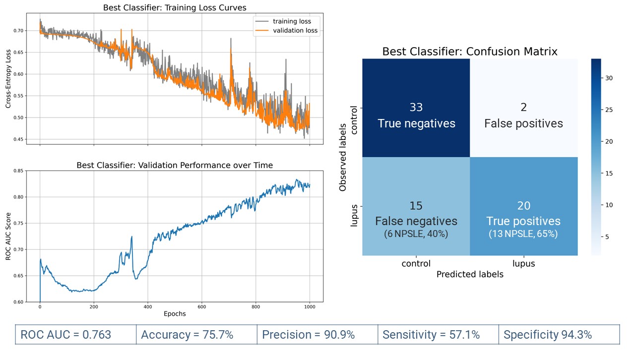
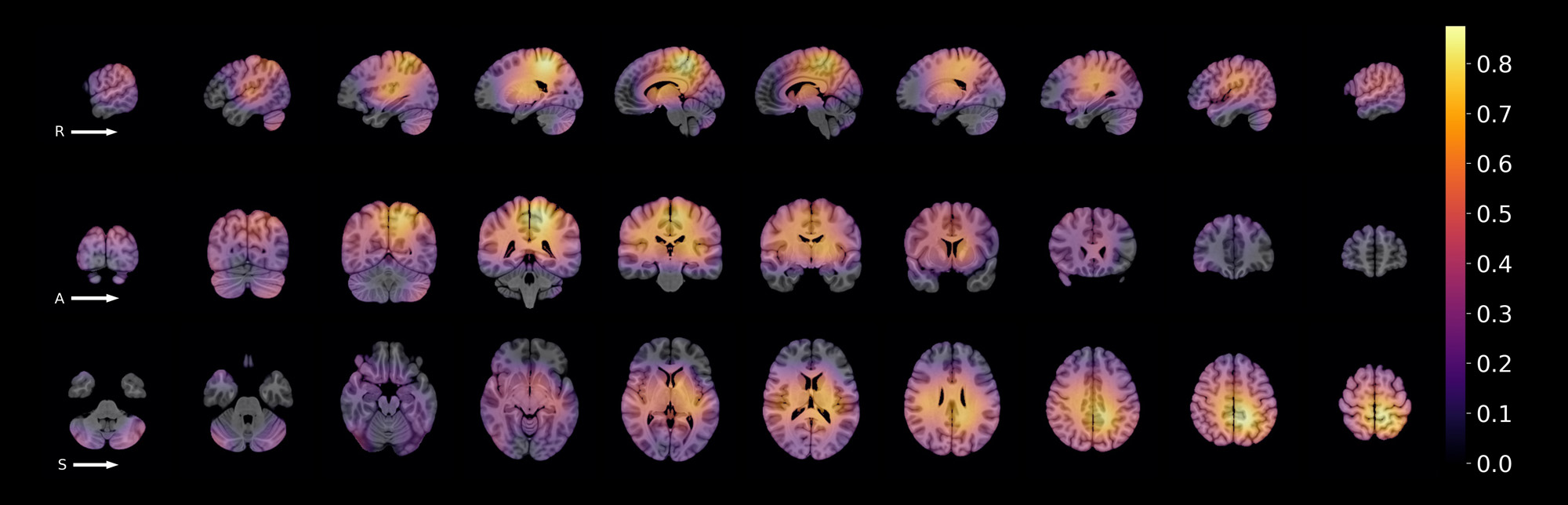
Results: Of all 233 cSLE images (133 patients), 127 (55%) were obtained from 57 patients with NPSLE. Most cSLE patients had mild/moderate disease activity at the time of scan (SLEDAI < = 10, n=153, 69.96%). When distinguishing cSLE vs non-cSLE, we achieved acceptable final classification performance with ROC AUC score = 0.763. From the 70 test samples (35 cSLE, 35 control), 20/35 of cSLE were correctly classified (true positives) (Fig. 2). The proportion of NPSLE samples in the true positives was 65%, and that in the false negatives, 40%. The difference between these proportions was not significant (z-score=1.47, p=0.07). The most important brain image features for this classification task were found in intra-ventricular, peri-ventricular, and left medial parietal regions.

Conclusion: The resulting model was effective in capturing structural brain MRI features distinguishing cSLE, with and without NPSLE, from non-cSLE patients. Our findings suggest the presence of regional brain abnormalities in cSLE patients without an NPSLE diagnosis. This model may serve as a novel diagnostic tool prototype, and as a hypothesis generating tool in future NPSLE biomarker research. Further study is needed to characterise brain image features for each type of NP manifestation observed in cSLE.

Supporting image 1

Figure 1. Methods summary for model training, configuration, evaluation, and interpretation. Grad-CAM stands for Gradient-weighted Class Activation Maps, a technique commonly used to interpret computer vision models.

Supporting image 2

Figure 2. Learning behaviour and final classification performance of the best model. The left panel illustrates normal learning behaviour through decreasing loss curves and increasing performance over time. The right panel contains a confusion matrix illustrating the classification performance of the model on a held-out test set at an optimal classification threshold of 0.60. The bottom table summarises final model performance through 5 different classification metrics.

Supporting image 3

Figure 3. A standard MNI brain overlaid with the average class activation map for the correctly classified cSLE test samples (n=20). Yellow and orange colours highlight image features of the brain that were consistently used to correctly classify samples as cSLE. The top row shows sagittal slices (left -> right); middle row shows coronal slices (anterior -> posterior), and bottom row shows axial slices (inferior -> superior).


Disclosures: s. arciniegas: None; M. Wagner: None; L. Vidarsson: None; B. Ertl-Wagner: None; T. El Tal: None; A. Jeyanathan: None; L. Ng: None; L. Hiraki: None; D. Levy: None; G. Ibrahim: LivaNova Inc., 1, 2, 5, Medtronic Inc., 2, Synergia Inc., 1, 2; A. Knight: Pfizer, 6.

To cite this abstract in AMA style:

arciniegas s, Wagner M, Vidarsson L, Ertl-Wagner B, El Tal T, Jeyanathan A, Ng L, Hiraki L, Levy D, Ibrahim G, Knight A. Identifying Potential Neuroimaging Biomarkers of Neuropsychiatric Lupus in Children Using Deep Learning [abstract]. Arthritis Rheumatol. 2023; 75 (suppl 9). https://acrabstracts.org/abstract/identifying-potential-neuroimaging-biomarkers-of-neuropsychiatric-lupus-in-children-using-deep-learning/. Accessed .
  • Tweet
  • Click to email a link to a friend (Opens in new window) Email
  • Click to print (Opens in new window) Print

« Back to ACR Convergence 2023

ACR Meeting Abstracts - https://acrabstracts.org/abstract/identifying-potential-neuroimaging-biomarkers-of-neuropsychiatric-lupus-in-children-using-deep-learning/

Advanced Search

Your Favorites

You can save and print a list of your favorite abstracts during your browser session by clicking the “Favorite” button at the bottom of any abstract. View your favorites »

All abstracts accepted to ACR Convergence are under media embargo once the ACR has notified presenters of their abstract’s acceptance. They may be presented at other meetings or published as manuscripts after this time but should not be discussed in non-scholarly venues or outlets. The following embargo policies are strictly enforced by the ACR.

Accepted abstracts are made available to the public online in advance of the meeting and are published in a special online supplement of our scientific journal, Arthritis & Rheumatology. Information contained in those abstracts may not be released until the abstracts appear online. In an exception to the media embargo, academic institutions, private organizations, and companies with products whose value may be influenced by information contained in an abstract may issue a press release to coincide with the availability of an ACR abstract on the ACR website. However, the ACR continues to require that information that goes beyond that contained in the abstract (e.g., discussion of the abstract done as part of editorial news coverage) is under media embargo until 10:00 AM ET on November 14, 2024. Journalists with access to embargoed information cannot release articles or editorial news coverage before this time. Editorial news coverage is considered original articles/videos developed by employed journalists to report facts, commentary, and subject matter expert quotes in a narrative form using a variety of sources (e.g., research, announcements, press releases, events, etc.).

Violation of this policy may result in the abstract being withdrawn from the meeting and other measures deemed appropriate. Authors are responsible for notifying colleagues, institutions, communications firms, and all other stakeholders related to the development or promotion of the abstract about this policy. If you have questions about the ACR abstract embargo policy, please contact ACR abstracts staff at [email protected].

Wiley

  • Online Journal
  • Privacy Policy
  • Permissions Policies
  • Cookie Preferences

© Copyright 2025 American College of Rheumatology