ACR Meeting Abstracts

ACR Meeting Abstracts

  • Meetings
    • ACR Convergence 2024
    • ACR Convergence 2023
    • 2023 ACR/ARP PRSYM
    • ACR Convergence 2022
    • ACR Convergence 2021
    • ACR Convergence 2020
    • 2020 ACR/ARP PRSYM
    • 2019 ACR/ARP Annual Meeting
    • 2018-2009 Meetings
    • Download Abstracts
  • Keyword Index
  • Advanced Search
  • Your Favorites
    • Favorites
    • Login
    • View and print all favorites
    • Clear all your favorites
  • ACR Meetings

Abstract Number: 0923

Identification of Lupus Nephritis Kidney Immune Populations via Hi-Resolution 20-Plex Immunohistochemistry Single-Cell Spatial Analyses

Caleb Marlin1, Alessandra Ida Celia2, Richard Furie3, Judith James1, Andrea Fava4, Joel Guthridge1, Avi Rosenberg5, Tayte Stephens1, Christian Wright1, Jeff Hodgin6, Peter Izmirly7, H Michael Belmont8, Jennifer Anolik9, Michelle Petri10, Jill Buyon11, Diane L. Kamen12, Chaim Putterman13, Accelerating Medicines Partnership in RA/SLE The1, Chen-Yu Lee4 and Xiaoping Yang4, 1Oklahoma Medical Research Foundation, Oklahoma City, OK, 2John Hopkins University of Medicine, Rome, Italy, 3Northwell Health, Manhasset, NY, 4Johns Hopkins University, Baltimore, MD, 5Department of Pathology, The Johns Hopkins University School of Medicine, Baltimore, MD, 6University of Michigan, Ann Arbor, MI, 7New York University School of Medicine, New York, NY, 8NYU School of Medicine, New York, NY, 9University of Rochester Medical Center, Rochester, NY, 10Department of Medicine, Division of Rheumatology, Johns Hopkins University School of Medicine, Timonium, MD, 11NYU Grossman School of Medicine, New York, NY, 12Medical University of South Carolina, Charleston, SC, 13Albert Einstein College of Medicine, Bronx, NY

Meeting: ACR Convergence 2023

Keywords: autoimmune diseases, Imaging, Lupus nephritis

  • Tweet
  • Click to email a link to a friend (Opens in new window) Email
  • Click to print (Opens in new window) Print
Session Information

Date: Monday, November 13, 2023

Title: (0899–0933) SLE – Etiology & Pathogenesis Poster

Session Type: Poster Session B

Session Time: 9:00AM-11:00AM

Background/Purpose: Immunohistochemistry is the gold standard for antibody staining in microscopy. However, a lack of multiplexing limits its utility in the use of discovery. Here we demonstrate a serial Immunohistochemistry (sIHC) staining workflow and analysis pipeline allowing for multiplexing of 18 antibodies on a single section.

Methods: 29 FFPE block were included for preliminary analyses from the 127 clinically indicated kidney biopsies recruited as part of the Lupus nephritis (LN) Accelerating Medicines Partnership (AMP) project).

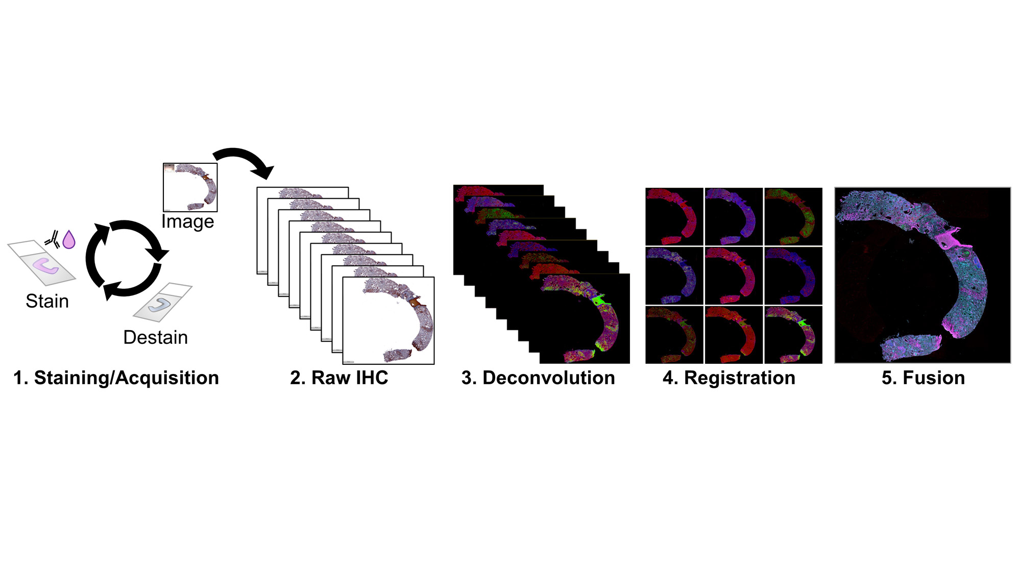
Staining: 10mm sections from LN kidney biopsies in FFPE were deparaffinized, rehydrated, and antigen retrieved with boiling citrate. Slides were blocked for peroxidases, washed, and incubated with primary antibody, secondary HRP reagents, AEC-Red Chromogen, and Hematoxylin. Images were acquired at 40X magnification. Slides were decolored in 90% Ethanol and stripped in an antibody elution buffer, then cycled for all markers.

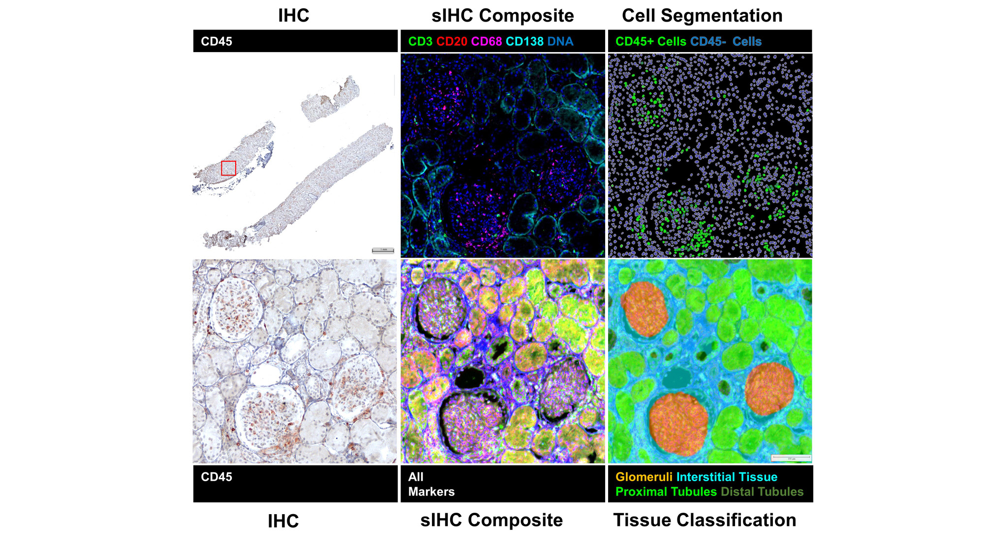
Image Stacks: Image files were imported into HALO-AI v3.5 (Indica Labs) for deconvolution, pseudo-coloring, co-registration, and alignment. Registered images were fused to create a single composite image with all 18 markers and a representative DNA and counterstain channel. Tissue regions were identified by a DenseNet V2 AI tissue classifier trained on an original co-registered IHC image and individual cell objects were identified by nuclear detection and segmented by cell distance boundaries.

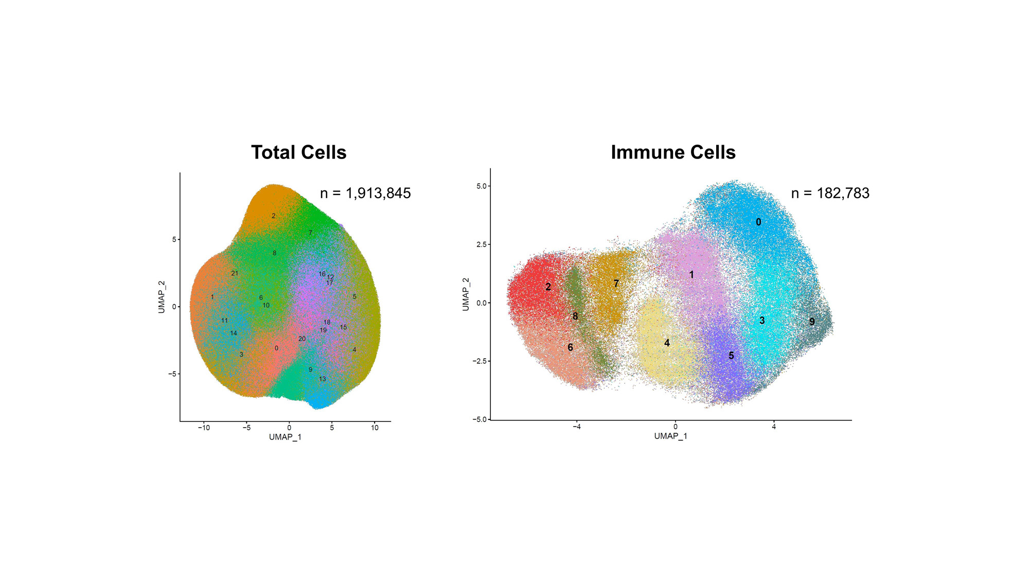
Analysis: Single-cell marker expression datasets were analyzed in R. Cells located on the edge of the tissue or in regions classified as wrinkles were removed to reduce potential artifacts. Marker expression values underwent CLR-normalization and standardization within each sample and then combined and harmonized by Harmony to further minimize batch effect. PCA and UMAP were used for dimensional reduction, and KNN and SNN algorithms were applied to identify cell clusters. Clusters with CD45 expression levels > 2 standard deviations were selected as candidate immune cell clusters. Cell clusters with high keratin marker expression were removed to eliminate renal tubular epithelial cells. Remaining clusters were then re-clustered via KNN and SNN to identify distinct immune cell types with the help of heatmaps and feature plots.

Results: The co-registration and fusion of images produced image stacks in which all 20 markers could be visualized within the same image. Tissue classification and single-cell segmentation allowed for clear identification of regions of the tissue and the individual cells that constituted each region. Single-cell analyses identified 1,913,845 cells and 182,783 CD45+ cells which were classified into 10 distinct immune cell phenotypes.

Conclusion: Here we demonstrate imaging and analysis of 20 markers using sIHC for the first time, while producing a large, spatially informed, clinically-relevant dataset in 29 LN Kidney Biopsies.sIHC can be successfully employed to perform multiplexed whole slide analysis harnessing both the subcellular resolution (brightfield) and the reliability of IHC.

Supporting image 1

Figure 1-sIHC Staining and Image Stack Pipeline

Supporting image 2

Figure 2 – sIHC Composites, Cell Segmentation, and AI-Based Tissue Classification

Supporting image 3

Figure 3 – Dimension Reduction and Clustering of sIHC Single-Cell Data


Disclosures: C. Marlin: None; A. Celia: None; R. Furie: Biogen, 2, 5; J. James: Bristol-Myers Squibb(BMS), 5, GlaxoSmithKlein(GSK), 2, Novartis, 2, Progentec Biosciences, 5; A. Fava: Annexon Biosciences, 2, Sanofi, 1; J. Guthridge: None; A. Rosenberg: None; T. Stephens: None; C. Wright: None; J. Hodgin: AstraZeneca, 5, 6, Eli Lilly, 5, Gilead, 5, Janssen, 5, Moderna, 5, Novo Nordisk, 5, Regeneron, 5; P. Izmirly: None; H. Belmont: Alexion, 6, Aurinia, 6; J. Anolik: None; M. Petri: Alexion, 1, Amgen, 1, AnaptysBio, 1, Annexon Bio, 1, Argenx, 1, Arhros-Focus Med/Ed, 6, AstraZeneca, 1, 5, Aurinia, 1, 5, 6, Axdev, 1, Biogen, 1, Boxer Capital, 2, Cabaletto Bio, 2, Caribou Biosciences, 2, CVS Health, 1, Eli Lilly, 1, 5, Emergent Biosolutions, 1, Exagen, 5, Exo Therapeutics, 2, Gilead Biosciences, 2, GlaxoSmithKlein(GSK), 1, 5, 6, Horizon Therapeutics, 2, Idorsia Pharmaceuticals, 2, IQVIA, 1, Janssen, 1, 5, Kira Pharmaceuticals, 2, MedShr, 6, Merck/EMD Serono, 1, Momenta Pharmaceuticals, 2, Nexstone Immunology, 2, Nimbus Lakshmi, 2, Proviant, 2, Sanofi, 2, Sinomab Biosciences, 2, Thermofisher, 5, UCB, 2; J. Buyon: Bristol-Myers Squibb(BMS), 2, GlaxoSmithKlein(GSK), 2, Related Sciences, 1; D. Kamen: None; C. Putterman: Equillium, 2, KidneyCure, 1, Progentec, 2; A. The: None; C. Lee: None; X. Yang: None.

To cite this abstract in AMA style:

Marlin C, Celia A, Furie R, James J, Fava A, Guthridge J, Rosenberg A, Stephens T, Wright C, Hodgin J, Izmirly P, Belmont H, Anolik J, Petri M, Buyon J, Kamen D, Putterman C, The A, Lee C, Yang X. Identification of Lupus Nephritis Kidney Immune Populations via Hi-Resolution 20-Plex Immunohistochemistry Single-Cell Spatial Analyses [abstract]. Arthritis Rheumatol. 2023; 75 (suppl 9). https://acrabstracts.org/abstract/identification-of-lupus-nephritis-kidney-immune-populations-via-hi-resolution-20-plex-immunohistochemistry-single-cell-spatial-analyses/. Accessed .
  • Tweet
  • Click to email a link to a friend (Opens in new window) Email
  • Click to print (Opens in new window) Print

« Back to ACR Convergence 2023

ACR Meeting Abstracts - https://acrabstracts.org/abstract/identification-of-lupus-nephritis-kidney-immune-populations-via-hi-resolution-20-plex-immunohistochemistry-single-cell-spatial-analyses/

Advanced Search

Your Favorites

You can save and print a list of your favorite abstracts during your browser session by clicking the “Favorite” button at the bottom of any abstract. View your favorites »

All abstracts accepted to ACR Convergence are under media embargo once the ACR has notified presenters of their abstract’s acceptance. They may be presented at other meetings or published as manuscripts after this time but should not be discussed in non-scholarly venues or outlets. The following embargo policies are strictly enforced by the ACR.

Accepted abstracts are made available to the public online in advance of the meeting and are published in a special online supplement of our scientific journal, Arthritis & Rheumatology. Information contained in those abstracts may not be released until the abstracts appear online. In an exception to the media embargo, academic institutions, private organizations, and companies with products whose value may be influenced by information contained in an abstract may issue a press release to coincide with the availability of an ACR abstract on the ACR website. However, the ACR continues to require that information that goes beyond that contained in the abstract (e.g., discussion of the abstract done as part of editorial news coverage) is under media embargo until 10:00 AM ET on November 14, 2024. Journalists with access to embargoed information cannot release articles or editorial news coverage before this time. Editorial news coverage is considered original articles/videos developed by employed journalists to report facts, commentary, and subject matter expert quotes in a narrative form using a variety of sources (e.g., research, announcements, press releases, events, etc.).

Violation of this policy may result in the abstract being withdrawn from the meeting and other measures deemed appropriate. Authors are responsible for notifying colleagues, institutions, communications firms, and all other stakeholders related to the development or promotion of the abstract about this policy. If you have questions about the ACR abstract embargo policy, please contact ACR abstracts staff at [email protected].

Wiley

  • Online Journal
  • Privacy Policy
  • Permissions Policies
  • Cookie Preferences

© Copyright 2025 American College of Rheumatology