Session Information
Session Type: ACR Poster Session B
Session Time: 9:00AM-11:00AM
Background/Purpose: Using the tumor necrosis factor transgenic (TNF-Tg) mouse model of rheumatoid arthritis (RA), we have shown that during progression of knee synovitis, popliteal lymph nodes (PLNs) initially expand and then collapse concomitant with arthritic flare in the ipsilateral knee. In these collapsed PLNs, there is an abundance of B220+/CD23+/CD21hi B cells in inflamed lymph nodes (Bin) clogging the lymph node sinuses. We have also recently shown that TNF-Tg mice have evidence of interstitial lung disease (ILD), with large numbers of B cells. To determine the relationship between these B cells and Bin, we performed histology to phenotype B-cells in PLN versus mediastinal lymph nodes (MLN) from WT and TNF-Tg mice.
Methods: MLNs and PLNs (n=3) were harvested from 12-month old male TNF-Tg mice (3647 line in C57B6 background) with established arthritis and ILD, and their WT littermates (MLN n=3, PLN n=6). Tissues were processed for immunohistochemistry with antibodies against B220, a pan B-cell marker, and DAPI and imaged at 10 and 20x magnifications. Fluorescent images were quantified by manually counting the number of high intensity areas in three 10x fields of view on each slide. Two slides per tissue type per animal were used, with six total images averaged for one count per tissue type per animal.
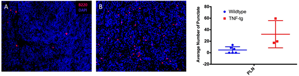
Results: PLNs from both WT and TNF-Tg animals showed a distinct pattern of increased high intensity staining compared to MLNs (4.74 vs 0 for WT, p<0.05 and 32.16 vs 0.36, p<0.05 for TNF-Tg). Furthermore, TNF-Tg PLNs showed increased high intensity staining compared to WT PLNs (32.16 vs 4.74, p<0.05) (Fig. 1).
Conclusion: This is the first description of a B220hi population in PLNs of aged mice with established arthritis and ILD. Interestingly, only PLNs display an increased B220hi population compared to MLNs, which drain the inflamed lung. The discordant expression in MLN versus PLN tissue suggests a continuous localized response of B cells in lymph nodes draining synovial tissue. Further specific characterization of this population at earlier timepoints during arthritis development is ongoing in order to determine if expression is altered in early disease, and if B220 expression pattern could help differentiate early inflammation from established disease. Figure 1: Representative B220hi images at 20x magnification of WT (A) and TNF-Tg (B) PLN
 |
To cite this abstract in AMA style:
Forney M, Bell R, Schwarz E, Rahimi H. Identification of a Unique Population of B220hi B-Cells in Inflamed Lymph Nodes (Bin) As a Potential Biomarker of Arthritic Progression in the Tumor Necrosis Factor Transgenic Mouse Model of Rheumatoid Arthritis [abstract]. Arthritis Rheumatol. 2016; 68 (suppl 10). https://acrabstracts.org/abstract/identification-of-a-unique-population-of-b220hi-b-cells-in-inflamed-lymph-nodes-bin-as-a-potential-biomarker-of-arthritic-progression-in-the-tumor-necrosis-factor-transgenic-mouse-model-of-rheumatoi/. Accessed .« Back to 2016 ACR/ARHP Annual Meeting
ACR Meeting Abstracts - https://acrabstracts.org/abstract/identification-of-a-unique-population-of-b220hi-b-cells-in-inflamed-lymph-nodes-bin-as-a-potential-biomarker-of-arthritic-progression-in-the-tumor-necrosis-factor-transgenic-mouse-model-of-rheumatoi/