Session Information
Date: Wednesday, November 8, 2017
Title: Metabolic and Crystal Arthropathies II: Mechanisms of Crystal Inflammation and Metabolism
Session Type: ACR Concurrent Abstract Session
Session Time: 9:00AM-10:30AM
Background/Purpose: Monosodium urate (MSU) crystal deposition and gout flares frequently affect joints that have been damaged or are affected by osteoarthritis. The aim of this study was to examine the effects of healthy, osteoarthritic and degraded human cartilage on the three key steps of MSU crystallization; reduced urate solubility, crystal nucleation (appearance of MSU crystals), and crystal growth.
Methods: Paired macroscopically-healthy and macroscopically-diseased knee cartilage samples were obtained from patients undergoing orthopedic surgery. Cartilage was homogenized using the FreezerMill 6870 (SPEX CertiPrep Ltd) and the resulting powder dispersed in sterile water for preparation of cartilage homogenates. In crystallization assays, cartilage homogenates were added to solutions of sodium urate at 37°C and pH adjusted to 8.9. The cartilage-urate solutions were sampled over 24 hours for assessment of urate concentration (Cobas c311 autoanalyser, Roche Diagnostics), and crystal growth and morphology (by polarizing microscopy and ImageJ software). After 24 hours, total MSU crystal weight was determined. In separate assays, time to nucleation was determined by polarizing microscopy. The following cartilage preparations were tested: 1%, 5% and 10% healthy homogenized cartilage; 5% healthy and 5% diseased homogenized cartilage; and 5% healthy homogenized cartilage and 5% healthy enzyme-degraded cartilage.
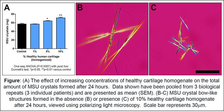
Results: The addition of 5% and 10% healthy cartilage homogenates increased the total weight of MSU crystals formed compared to control (no added cartilage) (1-way ANOVA, P=0.0007, Figure A). Addition of healthy cartilage did not change urate solubility or time to nucleation compared to control. MSU crystals grown in the presence of healthy cartilage homogenates were significantly shorter (2-way ANOVA, P<0.0001). In morphological analysis, MSU crystal bow-like structures were observed both in the presence and absence of healthy cartilage homogenates, and the bows formed in the presence of healthy cartilage were also significantly shorter compared to control (Figure B and C). There were no differences between healthy cartilage and diseased cartilage homogenates in all assessments. For the enzyme-degraded cartilage, the lengths of MSU crystals and MSU crystal bows grown in the presence of degraded cartilage were significantly shorter than those grown in the presence of homogenized cartilage (2-way ANOVA, P<0.0001 for both), with no difference in other measures.
Conclusion: In crystallization assays, addition of human cartilage increases the amount of MSU crystals formed and also influences MSU crystal size. These findings may provide an explanation for the clinical presentation of gout in osteoarthritic joints.
To cite this abstract in AMA style:
Chhana A, Pool B, Choi A, Gao R, Zhu M, Cornish J, Munro J, Dalbeth N. Human Cartilage Influences the Crystallization of Monosodium Urate; Understanding the Link between Gout and Osteoarthritis [abstract]. Arthritis Rheumatol. 2017; 69 (suppl 10). https://acrabstracts.org/abstract/human-cartilage-influences-the-crystallization-of-monosodium-urate-understanding-the-link-between-gout-and-osteoarthritis/. Accessed .« Back to 2017 ACR/ARHP Annual Meeting
ACR Meeting Abstracts - https://acrabstracts.org/abstract/human-cartilage-influences-the-crystallization-of-monosodium-urate-understanding-the-link-between-gout-and-osteoarthritis/

