ACR Meeting Abstracts

ACR Meeting Abstracts

  • Home
  • Meetings Archive
    • ACR Convergence 2022
    • ACR Convergence 2021
    • ACR Convergence 2020
    • 2020 ACR/ARP PRSYM
    • 2019 ACR/ARP Annual Meeting
    • 2018 ACR/ARHP Annual Meeting
    • 2017-2009 Meetings
    • Download Abstracts
  • Keyword Index
  • Advanced Search
  • Your Favorites
    • Favorites
    • Login
    • View and print all favorites
    • Clear all your favorites
  • Meeting Resource Center

Abstract Number: 0061

Harnessing Spatially-Resolved Gene Expression to Characterize the Transcriptional Landscape of Psoriatic Skin

Rochelle Castillo1, Ikjot Sidhu1, Di Yan1, Piotr Konieczny1, Rebecca Haberman1, Brandon Hsieh1, Andrea Neimann1, Shruti Naik1 and Jose Scher2, 1NYU Langone Health, New York, NY, 2New York University School of Medicine, New York, NY

Meeting: ACR Convergence 2021

Keywords: Gene Expression, Genomics and Proteomics, Psoriatic arthritis, RNA, skin

  • Tweet
  • Email
  • Print
Session Information

Date: Saturday, November 6, 2021

Session Title: Spondyloarthritis Including PsA – Basic Science Poster (0046–0068)

Session Type: Poster Session A

Session Time: 8:30AM-10:30AM

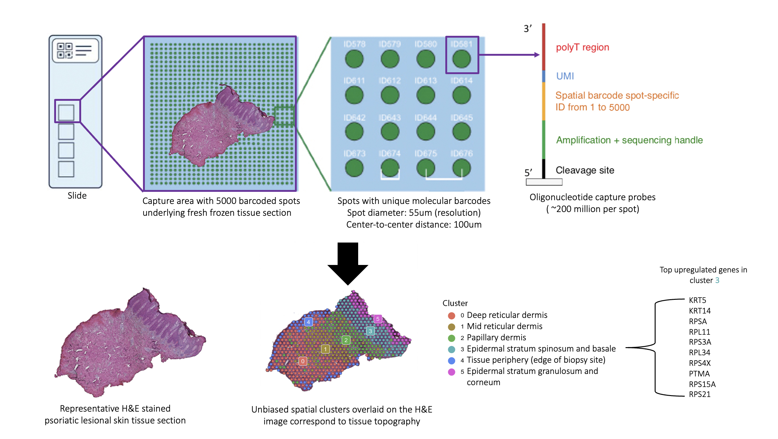
Background/Purpose: The skin is recognized as a window into the immunopathogenic mechanisms in the psoriatic arthritis (PsA) joint. This is evidenced by the fact that skin disease precedes joint involvement in ~90% of PsA patients and by the greater degree of overlap between gene expression in the synovial tissue of PsA and lesional skin in psoriasis (PsO) compared to synovium in other forms of inflammatory arthritis.1 Thus, the study of the psoriatic skin transcriptome has the potential to yield revolutionary insights into the immunopathogenesis of PsA. Spatial transcriptomics (ST) is a ground-breaking technology that allows for mRNA sequencing from histologically-intact tissue sections, facilitating precise localization of the site of gene expression (Fig. 1). A platform for ST (Visium Spatial Gene Expression Solution by 10X Genomics) has been made available; however, it remains to be optimized for human skin tissue, which is inherently challenging to use in transcriptomic studies due in part to its preponderance of RNAses.2,3 To date, there are no published studies on ST in either healthy or psoriatic human skin. Through a multidisciplinary collaboration, we have successfully optimized both healthy and psoriatic human skin tissue for use with ST.

Methods: Workflow

  1. Skin biopsy
  2. Cryopreservation
  3. Sectioning and staining
  4. Confirmation of RNA integrity
  5. Optimization of permeabilization
  6. Sequencing
  7. Data analysis

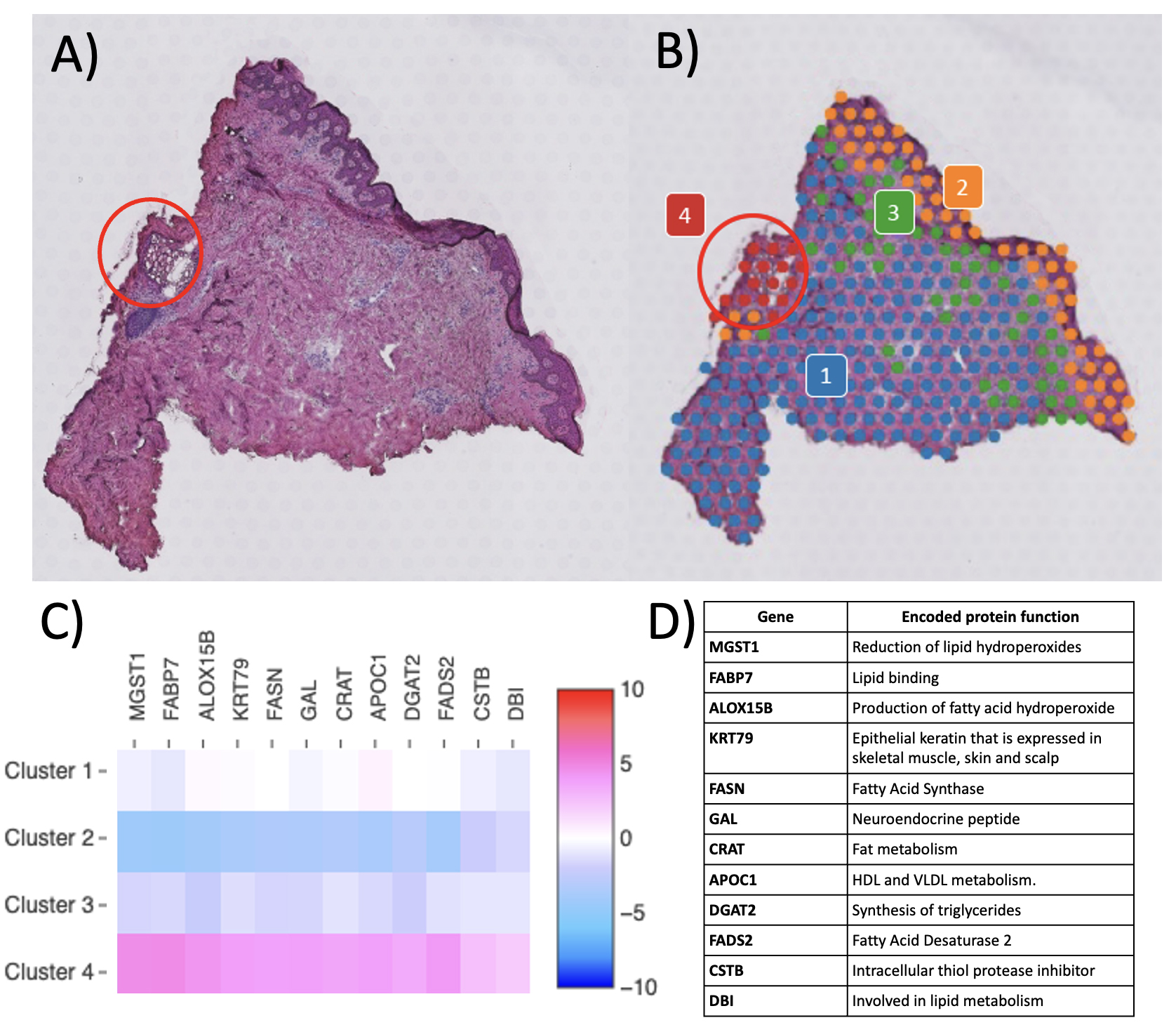
Results: To date, we have accrued samples from 3 controls, 4 PsA patients, and 6 PsO patients. All samples met the platform’s quality control (QC) metrics for spots and mapping, with reads in spots under tissue >50% (range: 63.1 to 87.1) and reads mapped confidently to exonic regions >30% (range: 80.1 to 91.5). The expected biological variation in transcriptional activity as evidenced by molecular counts (which correlate strongly with unique genes)4 across disease states and tissue regions was observed, with gene expression strikingly greater in the cell-dense epidermis and in appendageal structures than in the dermis and in psoriatic lesional skin compared to non-lesional and control skin (Fig. 2A). To detect technical variation, two contiguous sections from the same control sample were run on separate slides (Fig. 2B). Clustering and the Uniform Manifold Approximation and Projection (UMAP) plot architecture were consistent between the two replicates. Importantly, accuracy of spatial localization of gene expression and biological consistency of unbiased clustering was observed, with concordance of histopathologically annotated regions with gene-expression based clustering (Fig. 3).

Conclusion: Successful optimization of both healthy and psoriatic human skin tissue for ST was achieved, with all samples meeting the platform’s QC metrics. The expected biological variance in transcriptional activity across tissue regions and disease states was noted, the quantity, quality, and location of reads was biologically consistent, and there was no technical variation between samples. Thus, spatial profiling of gene expression in psoriatic skin through spatial transcriptomics can be performed and has the potential to offer invaluable insights into the immunopathogenesis of psoriatic disease.

Fig. 1. Overview of spatial transcriptomics. The spatial transcriptomics platform utilizes a slide containing four capture areas, each with 5000 molecularly barcoded, spatially encoded spots over which an intact fresh frozen tissue section is placed, stained, imaged, and permeabilized. Permeabilization results in the release of mRNA transcripts from the tissue that are then captured by ~200 million oligonucleotide capture probes. The unique spatial barcode allows the transcripts to be mapped to their exact location in the tissue section. Imaging and RNA sequencing data are processed together, resulting in whole transcriptome gene expression mapped to the tissue image. Each generated spatial cluster represents unbiased grouping based on gene expression and should ideally correlate with tissue topography. Gene expression in each cluster can then be explored.

Fig. 2. Transcriptional activity and reproducibility of spatial transcriptomics in control (non-psoriatic) and psoriatic skin. A.) Molecular counts mapped to each spot across disease states (psoriatic lesional, non-lesional, control) and tissue regions. Across all disease states, the epidermis and appendageal structures (hair follicles, sebaceous glands) were found to be more transcriptionally active than the dermis, consistent with the much higher cell density in these regions compared to the dermis, which consists mostly of collagen and elastic fibers and ground substance. Lesional skin from PsO and PsA patients was strikingly more transcriptionally active than non-lesional skin and control skin, which is consistent with epidermal hyperproliferation characteristic of psoriatic disease. B.) Determining reproducibility across runs. To detect technical variation, two contiguous sections from the same control sun-exposed skin sample (center and right panel) were run on the ST platform on separate slides. For comparison, results from a separate control sample are also shown (left panel). All three sections were computationally merged to render the clusters comparable. Identical clusters (middle row) and largely similar overall UMAP plot architecture (bottom row) was noted, save for an increase in the density of spots in the second run, which can be attributed to an increase in the volume of tissue overlying the spots. Of note, cluster 8, which contains the hair follicle (encircled in red), does not appear in the control sun-protected sample, which is devoid of hair follicles, and supports accuracy of spatial localization of gene expression.

Fig. 3. Concordance of histopathologic annotation with unbiased gene expression-based clustering. A.) H&E stained non-lesional skin section with sebaceous gland manually annotated (encircled in red). B.) Unbiased clustering of spots by gene expression results in unique segregation of sebaceous gland region (Cluster 4) (k means=4). C.) Heatmap of top globally differentially expressed genes (DEG) in the cluster containing the sebaceous glands (Cluster 4). D.) Description of DEG in cluster containing the sebaceous glands. The vast majority are involved in the metabolism of various lipids such as cholesterol, fatty acids, triglycerides which make up sebum, the production and secretion of which is the primary function of sebaceous glands.


Disclosures: R. Castillo, None; I. Sidhu, None; D. Yan, None; P. Konieczny, None; R. Haberman, Janssen, 1; B. Hsieh, None; A. Neimann, BMS, 12, Advisory Board, Celgene, 12, Advisory Board, Abbot, 11, Abbvie, 11, janssen, 11, Pfizer, 11; S. Naik, Seed Health, Inc, 1; J. Scher, Janssen, 2, 5, Novartis, 2, 5, Pfizer, 2, 5, AbbVie, 2, Sanofi, 2, Kaleido, 2, UCB, 2.

To cite this abstract in AMA style:

Castillo R, Sidhu I, Yan D, Konieczny P, Haberman R, Hsieh B, Neimann A, Naik S, Scher J. Harnessing Spatially-Resolved Gene Expression to Characterize the Transcriptional Landscape of Psoriatic Skin [abstract]. Arthritis Rheumatol. 2021; 73 (suppl 9). https://acrabstracts.org/abstract/harnessing-spatially-resolved-gene-expression-to-characterize-the-transcriptional-landscape-of-psoriatic-skin/. Accessed February 2, 2023.
  • Tweet
  • Email
  • Print

« Back to ACR Convergence 2021

ACR Meeting Abstracts - https://acrabstracts.org/abstract/harnessing-spatially-resolved-gene-expression-to-characterize-the-transcriptional-landscape-of-psoriatic-skin/

Advanced Search

Your Favorites

You can save and print a list of your favorite abstracts during your browser session by clicking the “Favorite” button at the bottom of any abstract. View your favorites »

ACR Pediatric Rheumatology Symposium 2020

© COPYRIGHT 2023 AMERICAN COLLEGE OF RHEUMATOLOGY

Wiley

  • Home
  • Meetings Archive
  • Advanced Search
  • Meeting Resource Center
  • Online Journal
  • Privacy Policy
  • Permissions Policies
  • Cookie Preferences