Session Information
Session Type: Poster Session B
Session Time: 9:00AM-11:00AM
Background/Purpose: Over one-third of people will develop knee osteoarthritis within 10 years of an anterior cruciate ligament (ACL) injury. Ultrasound may be used to monitor early pathological cartilage changes that occur after an ACL injury. However, it is unclear if a non-invasive quantitative ultrasound assessment correlates with arthroscopic cartilage damage. The purpose of our study was to compare quantitative cartilage ultrasound metrics between people with and without arthroscopic cartilage damage following an ACL injury.
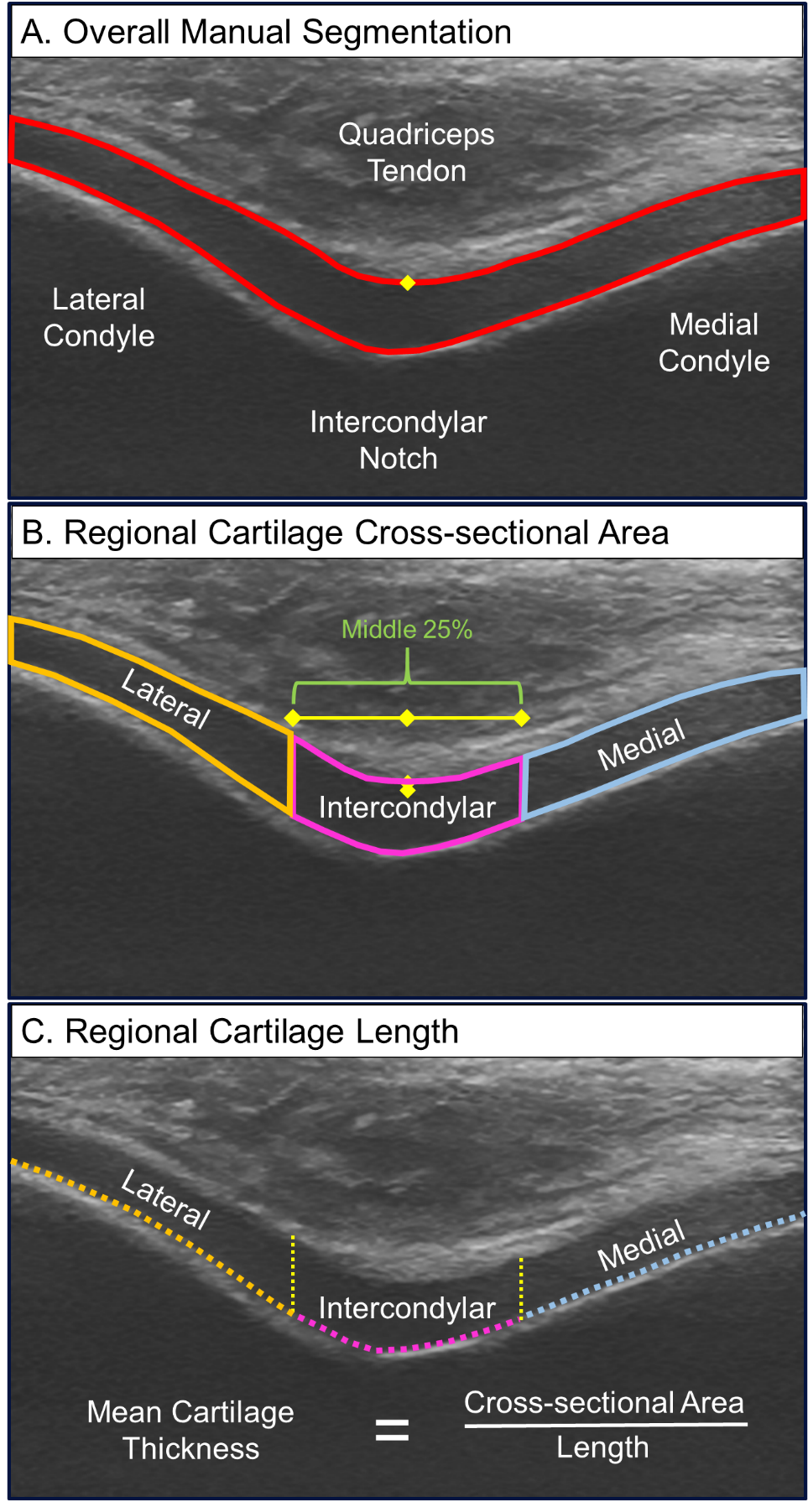
Methods: We recruited individuals between 18 and 35 years of age with a primary unilateral ACL injury at a pre-operative visit with an orthopaedic surgeon. Participants were excluded if they had a history of lower extremity surgery, injured either knee within the last 6 months (other than ACL injury), or previously been diagnosed with any form of arthritis. A knee ultrasound assessment occurred pre-operatively prior to an arthroscopy. With the knee in maximal knee flexion, a transverse suprapatellar ultrasound assessment was used to image the femoral cartilage in the individual’s injured knee. One reader manually segmented the cartilage. Next, a custom program automatically divided the cartilage into standardized medial and lateral femoral condyle regions and calculated 3 measures (Figure): 1) mean cartilage thickness (cartilage cross-sectional area divided by the cartilage length), 2) echo-intensity mean (average grey-scale pixel value ranging from 0–255), and 3) echo-intensity heterogeneity (standard deviation of the pixel intensity within the region). During a clinical arthroscopy, an orthopaedic surgeon with a sub-specialty in sports medicine graded the medial and femoral condyle using the Outerbridge grading system and operationally defined cartilage status as: normal cartilage: Outerbridge = 0 or cartilage damage: Outerbridge >1 (i.e., Outerbridge 1 = cartilage softening and swelling). We performed independent samples t-tests and used Cohen’s d effect sizes to compare the quantitative ultrasound metrics between individuals with (n=12) and without (n=12) arthroscopic cartilage damage.
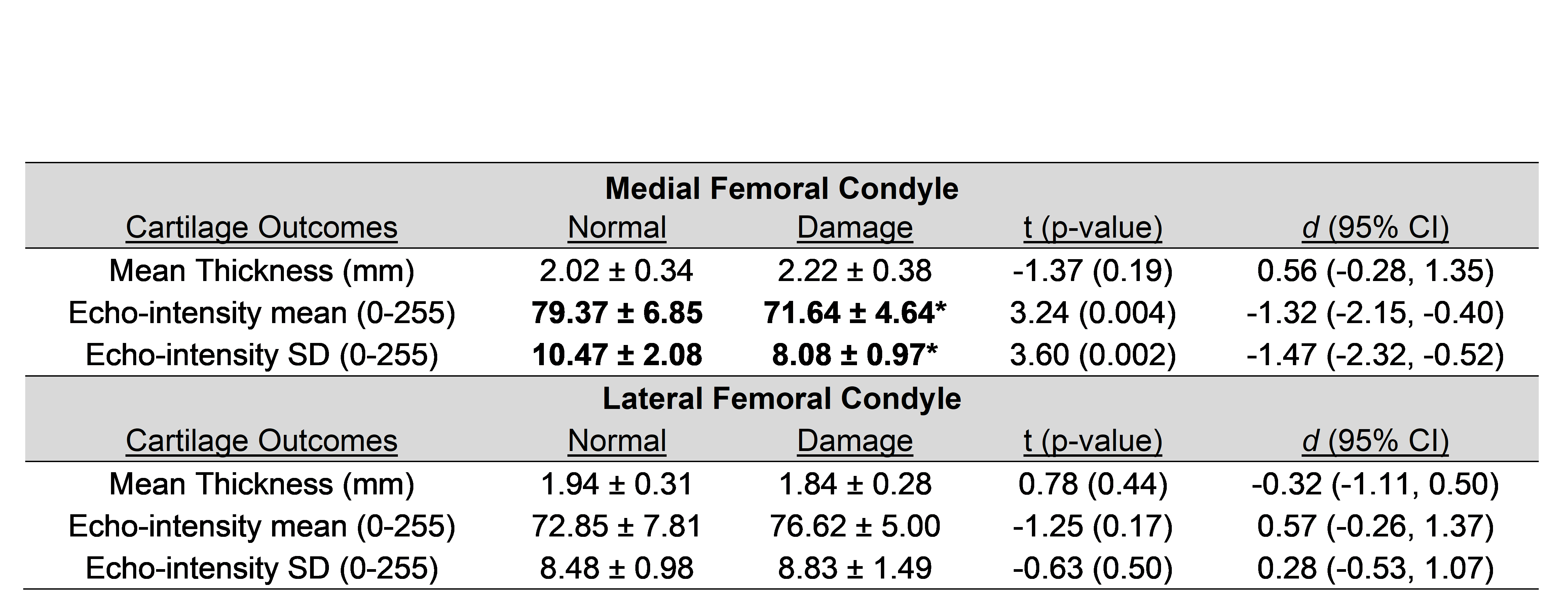
Results: The 24 participants were primarily male (n=16) and on average 24 + 5 years old and 50 + 52 days since ACL injury (Table 1). In the medial femoral condyle, echo-intensity mean and heterogeneity are greater in individuals with normal cartilage compared to those with cartilage damage (Table 2). In the lateral femoral condyle, there are no differences between cartilage ultrasound outcomes between individuals with and without arthroscopic cartilage damage.
Conclusion: Following ACL injury, an ultrasound assessment detects hypo-intense and less heterogeneous medial femoral cartilage in people with arthroscopically identified medial femoral cartilage damage. Future studies are needed to identify the prognostic importance of cartilage ultrasound echo-intensity characteristics, and validate these clinically-accessible metrics against more sophisticated markers of cartilage health.
 Table 2. Comparison of Femoral Cartilage Ultrasound Outcomes Between People With and Without Arthroscopic Cartilage Damage. SD = standard deviation; d = Cohen’s d effect size; 95% CI = 95% confidence intervals. *statistically significant compared to people with arthroscopically normal cartilage; Bold text indicates statistically significant and large magnitude effect sizes between people with and without cartilage damage
 Figure. Standardized Femoral Cartilage Segmentation. We used a custom program to take an overall cartilage manual segmentation (A), and use a standardized method to automatically separate the cartilage (B). This abstract focused on cartilage in the medial and lateral regions. The custom program calculated the mean cartilage thickness by dividing the regional cartilage cross-sectional area (B) by the regional cartilage length (C).
To cite this abstract in AMA style:
Harkey M, Little E, Thompson M, Zhang M, Driban J, Salzler M. Femoral Cartilage Ultrasound Echo-Intensity Associates with Arthroscopic Cartilage Damage in People Following Anterior Cruciate Ligament Reconstruction [abstract]. Arthritis Rheumatol. 2020; 72 (suppl 10). https://acrabstracts.org/abstract/femoral-cartilage-ultrasound-echo-intensity-associates-with-arthroscopic-cartilage-damage-in-people-following-anterior-cruciate-ligament-reconstruction/. Accessed .« Back to ACR Convergence 2020
ACR Meeting Abstracts - https://acrabstracts.org/abstract/femoral-cartilage-ultrasound-echo-intensity-associates-with-arthroscopic-cartilage-damage-in-people-following-anterior-cruciate-ligament-reconstruction/

