Session Information
Session Type: Poster Session C
Session Time: 10:30AM-12:30PM
Background/Purpose: Juvenile dermatomyositis (JDM) is a rare pediatric autoimmune myopathy characterized by skin and muscle inflammation, resulting in microvascular changes that can be visualized in the nailfold capillaries. Nailfold video capillaroscopy (NVC) at 200x magnification is a promising non-invasive measure to study disease activity. Here, we studied the association of nailfold capillary measurements with disease activity and compared rheumatologists’ examination (RE) at 10x magnification to NVC.
Methods: Patients diagnosed with JDM at a single tertiary care pediatric rheumatology center had nailfold capillaries imaged during clinic visits following published protocols. Clinical data (e.g., skin assessments and visual analog scale (VAS) scores) and notes on capillary appearances were extracted from medical records. Two cohorts were defined based on disease activity status at time of imaging: active JDM (physician VAS score > 0.5) and inactive JDM (physician VAS score < 0.5). Automated analysis of the NVC images were completed by Capillary.io, measuring capillary limb widths and apical diameters, followed by manual review for accuracy, apex widths, and internal diameters (Table 2, Fig 1). Each capillary characteristic was averaged per patient before analysis. Fisher and Wilcoxon rank sum tests were applied for categorical and continuous variables respectively. Data was stratified by RE at 10x: normal, mild-minimal, and abnormal. One-way ANOVA on ranks was applied, to assess for differences across the groups (P-value < 0.01). NVC measurements were compared to a published study of healthy pediatric capillaries.
Results: 29 JDM patients (mean age 11.79 ± 5.81) had 169 images analyzed, divided into 2 groups of 14 active and 15 inactive patients. Analysis of clinical variables between the two cohorts revealed active JDM exhibited more active skin disease and presence of periungual capillary loop change, active calcinosis, malar erythema, and chronic muscle weakness (Table 1). Active JDM was associated with decreased capillary density and enlarged arterial limb widths, apex widths, and internal diameters on NVC (Table 2, Fig 1). There was no significant difference in abnormal morphology between the two groups, but inactive JDM had significantly more normal capillaries per millimeter (Table 2). Between 80% to 100% of inactive JDM had NVC measurements within normal ranges, compared to about half of those with active JDM. Analysis of RE revealed patients described as “normal” on 10x met 4-5 of the normal 200x ranges, while “abnormal” met none.
Conclusion: Abnormal capillaries observed at 10x and 200x magnification were associated with active skin disease and muscle weakness in JDM, suggesting that nailfold capillaries, specifically arterial limb and apex widths, may serve as a clinical biomarker of disease activity. In addition, NVC showed abnormal measures could be detected and quantified in patients described clinically as “abnormal” or “mild-minimal” changes. Determining the clinical utility added by NVC will be an important area of future study along with identifying features that can help predict future flares, remission, and guide medications tapering.
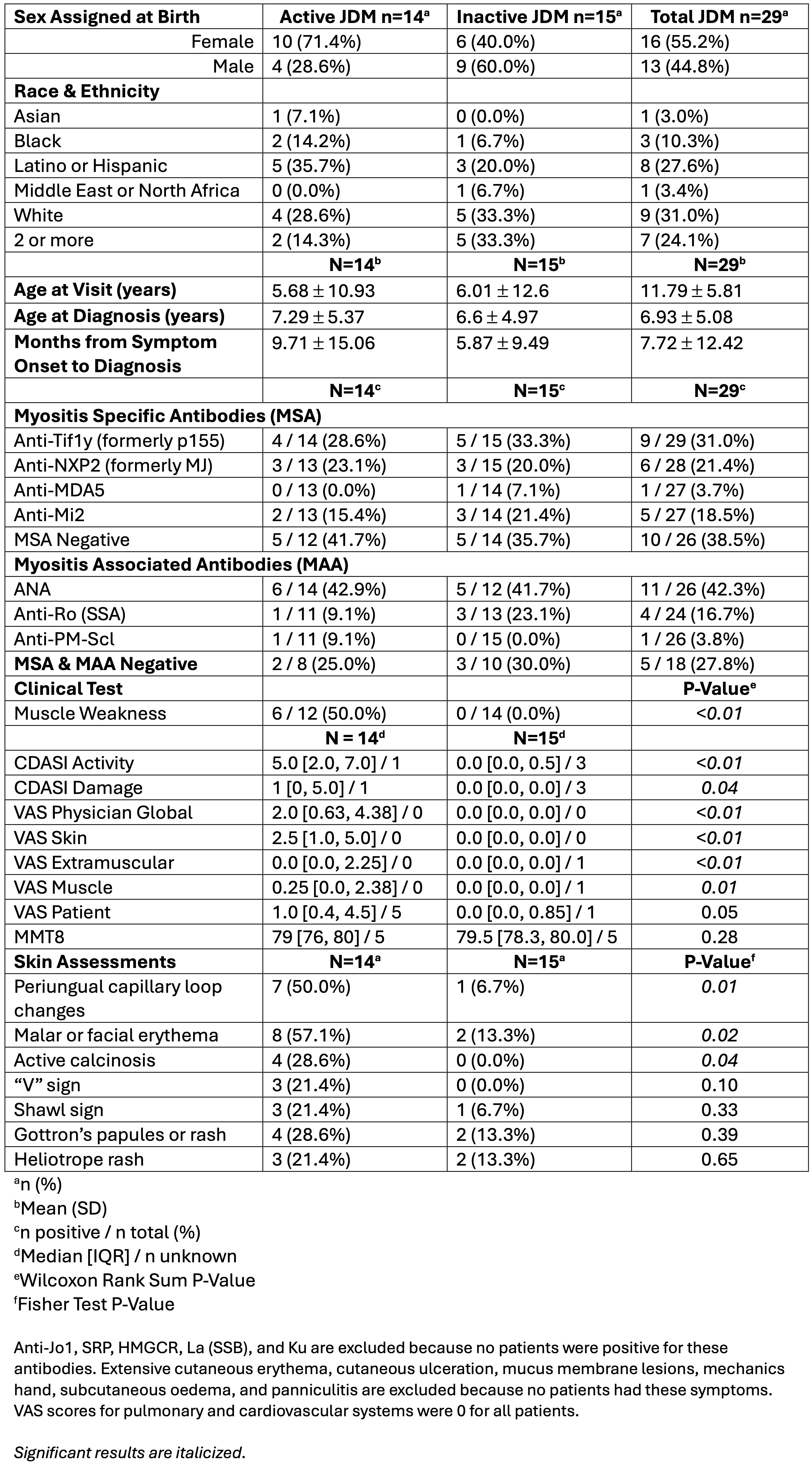 Table 1. Demographics & Disease Activity of Patients with Juvenile Dermatomyositis (JDM) with Nailfold Capillary imaging
.jpg) Table 2. Analysis of capillaries
Table 2. Analysis of capillaries
.jpg) Figure 1. “Active” versus “Inactive” JDM Capillaries
Figure 1. “Active” versus “Inactive” JDM Capillaries
To cite this abstract in AMA style:
Zheng Z, Metni L, Kim S, Neely J. Evaluating Nailfold Capillary Changes as Indicators of Disease Activity in Juvenile Dermatomyositis [abstract]. Arthritis Rheumatol. 2025; 77 (suppl 9). https://acrabstracts.org/abstract/evaluating-nailfold-capillary-changes-as-indicators-of-disease-activity-in-juvenile-dermatomyositis/. Accessed .« Back to ACR Convergence 2025
ACR Meeting Abstracts - https://acrabstracts.org/abstract/evaluating-nailfold-capillary-changes-as-indicators-of-disease-activity-in-juvenile-dermatomyositis/