ACR Meeting Abstracts

ACR Meeting Abstracts

  • Meetings
    • ACR Convergence 2024
    • ACR Convergence 2023
    • 2023 ACR/ARP PRSYM
    • ACR Convergence 2022
    • ACR Convergence 2021
    • ACR Convergence 2020
    • 2020 ACR/ARP PRSYM
    • 2019 ACR/ARP Annual Meeting
    • 2018-2009 Meetings
    • Download Abstracts
  • Keyword Index
  • Advanced Search
  • Your Favorites
    • Favorites
    • Login
    • View and print all favorites
    • Clear all your favorites
  • ACR Meetings

Abstract Number: 0874

Defining a Protective Role for Mucosal-associated Invariant T (MAIT) Cells in Spontaneous Cutaneous Lupus Erythematosus

Grace Crossland1, Lindsay Mendyka2, Michael Constantinides3 and Sladjana Skopelja-Gardner1, 1Dartmouth Geisel School of Medicine, Lebanon, NH, 2Dartmouth Hitchcock Memorial Hospital, Lyme, NH, 3Scripps Research Institute, San Diego, CA

Meeting: ACR Convergence 2024

Keywords: skin, T Cell, Treg cells

  • Tweet
  • Click to email a link to a friend (Opens in new window) Email
  • Click to print (Opens in new window) Print
Session Information

Date: Saturday, November 16, 2024

Title: Abstracts: T Cell Biology & Targets in Autoimmune & Inflammatory Disease

Session Type: Abstract Session

Session Time: 3:00PM-4:30PM

Background/Purpose: Systemic lupus erythematosus (SLE) is a leading cause of death in young females, and 70-85% of patients experience skin disease (cutaneous LE, CLE). The mechanisms of CLE pathogenesis remain unknown, and current treatments rely on broad immunosuppressive agents. Mucosal-associated invariant T (MAIT) cells, innate-like T cells that respond to microbial-derived antigens, have tissue repair capacity at homeostasis but are reduced in CLE skin. We hypothesize that restoring MAIT cell pools in non-lesional CLE-like skin will attenuate disease.

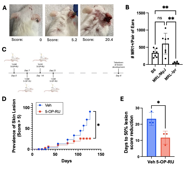
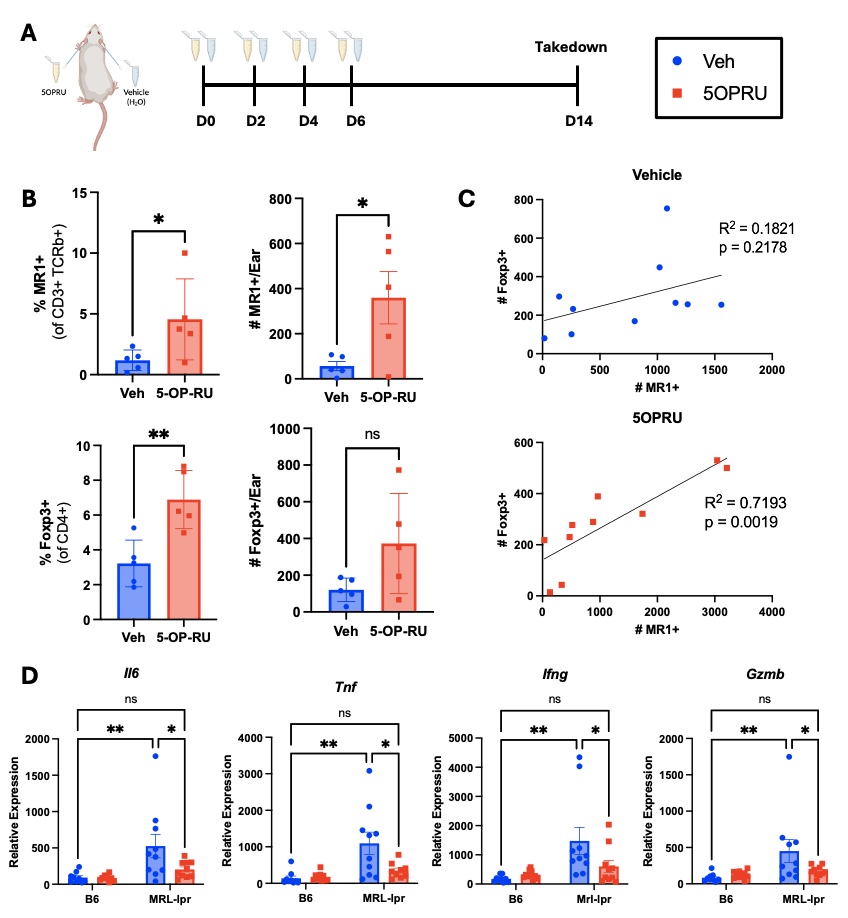
Methods: To test if expanding MAIT cells slows CLE development, female MRL-lpr mice were treated topically with the MAIT cell antigen 5-OP-RU (1mM) weekly starting at 8 weeks of age, and skin lesions were photographically documented and quantified by extent of erythema, scaling, thickness, and alopecia (Fig. 1A). To assess how MAIT cell expansion affects skin immune and inflammatory environment, 8-week-old female MRL-lpr mice were treated with 5-OP-RU topically every 48 hours for 4 total applications (Fig. 2). Skin immune and inflammatory environment was assessed by flow cytometry and qPCR.

Results: Analogous to the observations in the skin of lupus patients, MRL-lpr mice have fewer MAIT cells in non-lesional skin, compared to the control MRL/MpJ and B6 strains (Fig. 1B). Weekly topical application of the MAIT cell antigen 5-OP-RU, beginning prior to lesion development (Fig. 1C), reduced the prevalence of severe skin lesions compared to vehicle treated controls (Fig. 1D). Of the mice that did develop lesions, those that received 5-OP-RU displayed accelerated lesion healing (Fig. 1E). On average, 5-OP-RU-treated mice exhibited 50% reduction in lesion score 7 days faster than vehicle treated controls. 5-OP-RU topical application effectively expanded MAIT cells in Mrl-lpr skin (Fig. 2A). Surprisingly, MAIT expansion was accompanied by a concurrent increase in regulatory T cells (Treg) in the skin (Fig. 2B), and Treg numbers positively correlated with MAIT cell numbers (Fig. 2C). Moreover, MAIT cell expansion with 5-OP-RU reduced the high baseline expression of inflammatory (Il-6, Tnf) and cytotoxic (Ifng, Gzmb) genes in MRL-lpr skin, to the low levels of healthy B6 skin (Fig. 2D).

Conclusion: Topical application of the MAIT cell antigen 5-OP-RU slows development and progression of CLE-like lesions, suggesting a protective role for these cells in the skin. The concurrent expansion of Treg supports a potential immunosuppressive mechanism by which MAIT cells downregulate the expression of inflammatory and cytotoxic mediators in MRL-lpr skin associated with lesion development.

Supporting image 1

Figure 1. MAIT cell expansion attenuates lupus skin disease in MRL-lpr mice. A) Representative lesion scores quantified by extent of erythema, scaling, thickness, and alopecia. B) MAIT cell numbers in ear skin by genotype as determined by 5-OP-RU-loaded MR1 tetramer staining (n = 7-8 per group; One-way ANOVA, *p < 0.05, **p < 0.01). C) Experimental design for MAIT expansion via topical 5-OP-RU (1mM) in female MRL-lpr mice beginning at 8 weeks old. D) Lesion (score > 5) prevalence in 5-OP-RU and vehicle treated groups ( n = 4 per group; Log-rank test, *p < 0.05). E) Time to 50% lesion score reduction (Student’s t-test, n = 3 per group; *p < 0.05; one vehicle mouse died before 50% lesion resolution and one 5-OP-RU treated mouse never developed skin lesions, hence these are not included in the analysis in C).

Supporting image 2

Figure 2. MAIT cell expansion leads to Treg expansion and reduction in inflammatory mediators in non-lesional MRL-lpr skin. A) Experimental design for expansion of MAIT cells by topical 5-OP-RU (1mM) application in female 8 weeks old B6 and Mrl-lpr mice. B-C) MAIT (CD3+TCRb+gdTCR-MR1-5OPRU+) and Treg (CD4+Foxp3+) levels (percent and number) in the skin were quantified by flow cytometry at takedown. D) mRNA levels of inflammatory genes in B6 and Mrl-lpr skin, relative to Gapdh, with MAIT expansion (5-OP-RU, red) vs. vehicle (blue) quantified by qPCR at takedown. Statistical significance was determined by (B) Student’s t-test, (C) Pearson’s correlation analysis and (D) Two-way ANOVA with multiple comparisons; n=5_10 per group; *p < 0.05; **p < 0.01).


Disclosures: G. Crossland: None; L. Mendyka: None; M. Constantinides: None; S. Skopelja-Gardner: None.

To cite this abstract in AMA style:

Crossland G, Mendyka L, Constantinides M, Skopelja-Gardner S. Defining a Protective Role for Mucosal-associated Invariant T (MAIT) Cells in Spontaneous Cutaneous Lupus Erythematosus [abstract]. Arthritis Rheumatol. 2024; 76 (suppl 9). https://acrabstracts.org/abstract/defining-a-protective-role-for-mucosal-associated-invariant-t-mait-cells-in-spontaneous-cutaneous-lupus-erythematosus/. Accessed .
  • Tweet
  • Click to email a link to a friend (Opens in new window) Email
  • Click to print (Opens in new window) Print

« Back to ACR Convergence 2024

ACR Meeting Abstracts - https://acrabstracts.org/abstract/defining-a-protective-role-for-mucosal-associated-invariant-t-mait-cells-in-spontaneous-cutaneous-lupus-erythematosus/

Advanced Search

Your Favorites

You can save and print a list of your favorite abstracts during your browser session by clicking the “Favorite” button at the bottom of any abstract. View your favorites »

All abstracts accepted to ACR Convergence are under media embargo once the ACR has notified presenters of their abstract’s acceptance. They may be presented at other meetings or published as manuscripts after this time but should not be discussed in non-scholarly venues or outlets. The following embargo policies are strictly enforced by the ACR.

Accepted abstracts are made available to the public online in advance of the meeting and are published in a special online supplement of our scientific journal, Arthritis & Rheumatology. Information contained in those abstracts may not be released until the abstracts appear online. In an exception to the media embargo, academic institutions, private organizations, and companies with products whose value may be influenced by information contained in an abstract may issue a press release to coincide with the availability of an ACR abstract on the ACR website. However, the ACR continues to require that information that goes beyond that contained in the abstract (e.g., discussion of the abstract done as part of editorial news coverage) is under media embargo until 10:00 AM ET on November 14, 2024. Journalists with access to embargoed information cannot release articles or editorial news coverage before this time. Editorial news coverage is considered original articles/videos developed by employed journalists to report facts, commentary, and subject matter expert quotes in a narrative form using a variety of sources (e.g., research, announcements, press releases, events, etc.).

Violation of this policy may result in the abstract being withdrawn from the meeting and other measures deemed appropriate. Authors are responsible for notifying colleagues, institutions, communications firms, and all other stakeholders related to the development or promotion of the abstract about this policy. If you have questions about the ACR abstract embargo policy, please contact ACR abstracts staff at [email protected].

Wiley

  • Online Journal
  • Privacy Policy
  • Permissions Policies
  • Cookie Preferences

© Copyright 2025 American College of Rheumatology