ACR Meeting Abstracts

ACR Meeting Abstracts

  • Meetings
    • ACR Convergence 2024
    • ACR Convergence 2023
    • 2023 ACR/ARP PRSYM
    • ACR Convergence 2022
    • ACR Convergence 2021
    • ACR Convergence 2020
    • 2020 ACR/ARP PRSYM
    • 2019 ACR/ARP Annual Meeting
    • 2018-2009 Meetings
    • Download Abstracts
  • Keyword Index
  • Advanced Search
  • Your Favorites
    • Favorites
    • Login
    • View and print all favorites
    • Clear all your favorites
  • ACR Meetings

Abstract Number: 2590

Computer Vision Applied to Dual Energy Computed Tomography Images for Precise Calcinosis Cutis Quantification in Patients with Systemic Sclerosis

Anita Chandrasekaran1, Imran Omar 2, Zhicheng Fu 3, Shangping Ren 3 and Monique Hinchcliff 1, 1Yale University, Section of Rheumatology, Allergy and Immunology, New Haven, CT, 2Northwestern University Feinberg School of Medicine, Chicago, IL, 3San Diego State University, Department of Computer Science, San Diego, CA

Meeting: 2019 ACR/ARP Annual Meeting

Keywords: calcinosis, computed tomography (CT) and imaging techniques, Scleroderma, Systemic sclerosis

  • Tweet
  • Click to email a link to a friend (Opens in new window) Email
  • Click to print (Opens in new window) Print
Session Information

Date: Tuesday, November 12, 2019

Title: Systemic Sclerosis & Related Disorders – Clinical Poster III

Session Type: Poster Session (Tuesday)

Session Time: 9:00AM-11:00AM

Background/Purpose: Calcinosis cutis, found in both systemic sclerosis (SSc) and juvenile dermatomyositis patients, can be extensive and debilitating. Potential treatments have been identified, but a standardized and validated method for precise and accurate whole-body calcinosis cutis burden quantification is necessary to allow for valid clinical trials. Dual energy computed tomography (DECT) can differentiate between calcinosis cutis and healthy bone, but a radiologist must manually quantify the irregularly shaped lesions, which is time-consuming and costly. Computer vision, including convolutional neural network (CNN) algorithms, has been increasingly applied to solve problems in clinical medicine with success. The aim of this study is to optimize a CNN algorithm to facilitate quantification of calcinosis cutis disease burden.

Methods: De-identified 2-dimensional (2-D) DECT images from patients with SSc, with clinically apparent calcinosis cutis, were obtained. An expert musculoskeletal radiologist manually segmented the three forefinger phalanges to serve as the gold standard comparison. Computer scientists then trained and tested the CNN algorithm for finger bone segmentation. After reliable finger bone segmentation was achieved, the area of a calcinosis cutis lesion was measured and compared to measurements performed by a radiologist to test the utility of the CNN approach.

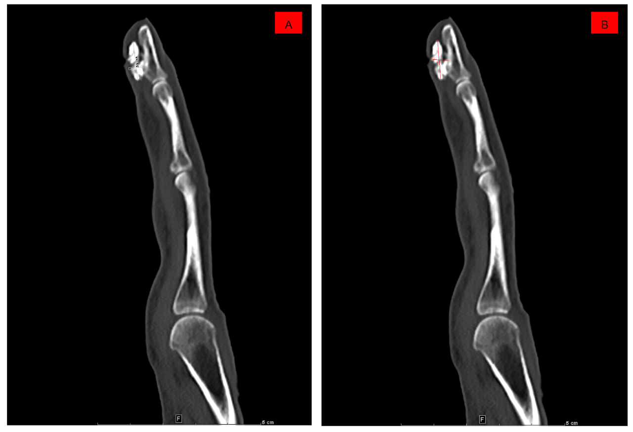
Results: Thirty 2D DECT hand images from patients with SSc were used to identify the most appropriate CNN algorithm for finger bone segmentation (Image 1A, representative image). The U-net CNN algorithm demonstrated superior performance. 500 epochs were necessary to adequately segment healthy finger bones from adjacent dystrophic calcifications (Image 1B). Calcinosis cutis lesions were then identified by subtracting normal finger bones from the image to permit the computer to “see” the dystrophic calcium lesions (Image 1C). When measured by a radiologist, the length x width of the lesion was used to calculate the 2-D area, assuming a relatively smooth lesion contour (Image 2A), whereas the CNN-calculated 2-D area utilized pixel intensities (Image 2B).

Conclusion: To our knowledge, the present study is the first to apply computer vision to the challenge of calcinosis cutis quantification. We demonstrate that CNN algorithms applied to DECT hand images of SSc patients can be used to differentiate calcinosis cutis lesions from adjacent healthy bone. When compared to the gold standard of radiologist review, CNN algorithms take into account lesion irregularity, which may allow for more precise measurements. Future work will involve lesion volume and density quantification, crucial for assessing response to treatment, as well as validation in a larger, independent cohort.

Image 1A: Representative 2D DECT hand image from a patient with SSc
Image 1B: Segmented healthy finger bone
Image 1C: Highlighted calcinosis cutis lesion, identified by subtracting normal finger bone from the overall image, emphasizing the area of dystrophic calcium

A demonstrates a radiologist-measured calcinosis cutis lesion, using the length x width of the lesion and assuming a relatively smooth contour, while B demonstrates the same lesion measured by the CNN algorithm, utilizing pixel intensities.


Disclosure: A. Chandrasekaran, None; I. Omar, None; Z. Fu, None; S. Ren, None; M. Hinchcliff, None.

To cite this abstract in AMA style:

Chandrasekaran A, Omar I, Fu Z, Ren S, Hinchcliff M. Computer Vision Applied to Dual Energy Computed Tomography Images for Precise Calcinosis Cutis Quantification in Patients with Systemic Sclerosis [abstract]. Arthritis Rheumatol. 2019; 71 (suppl 10). https://acrabstracts.org/abstract/computer-vision-applied-to-dual-energy-computed-tomography-images-for-precise-calcinosis-cutis-quantification-in-patients-with-systemic-sclerosis/. Accessed .
  • Tweet
  • Click to email a link to a friend (Opens in new window) Email
  • Click to print (Opens in new window) Print

« Back to 2019 ACR/ARP Annual Meeting

ACR Meeting Abstracts - https://acrabstracts.org/abstract/computer-vision-applied-to-dual-energy-computed-tomography-images-for-precise-calcinosis-cutis-quantification-in-patients-with-systemic-sclerosis/

Advanced Search

Your Favorites

You can save and print a list of your favorite abstracts during your browser session by clicking the “Favorite” button at the bottom of any abstract. View your favorites »

All abstracts accepted to ACR Convergence are under media embargo once the ACR has notified presenters of their abstract’s acceptance. They may be presented at other meetings or published as manuscripts after this time but should not be discussed in non-scholarly venues or outlets. The following embargo policies are strictly enforced by the ACR.

Accepted abstracts are made available to the public online in advance of the meeting and are published in a special online supplement of our scientific journal, Arthritis & Rheumatology. Information contained in those abstracts may not be released until the abstracts appear online. In an exception to the media embargo, academic institutions, private organizations, and companies with products whose value may be influenced by information contained in an abstract may issue a press release to coincide with the availability of an ACR abstract on the ACR website. However, the ACR continues to require that information that goes beyond that contained in the abstract (e.g., discussion of the abstract done as part of editorial news coverage) is under media embargo until 10:00 AM ET on November 14, 2024. Journalists with access to embargoed information cannot release articles or editorial news coverage before this time. Editorial news coverage is considered original articles/videos developed by employed journalists to report facts, commentary, and subject matter expert quotes in a narrative form using a variety of sources (e.g., research, announcements, press releases, events, etc.).

Violation of this policy may result in the abstract being withdrawn from the meeting and other measures deemed appropriate. Authors are responsible for notifying colleagues, institutions, communications firms, and all other stakeholders related to the development or promotion of the abstract about this policy. If you have questions about the ACR abstract embargo policy, please contact ACR abstracts staff at [email protected].

Wiley

  • Online Journal
  • Privacy Policy
  • Permissions Policies
  • Cookie Preferences

© Copyright 2025 American College of Rheumatology