ACR Meeting Abstracts

ACR Meeting Abstracts

  • Meetings
    • ACR Convergence 2024
    • ACR Convergence 2023
    • 2023 ACR/ARP PRSYM
    • ACR Convergence 2022
    • ACR Convergence 2021
    • ACR Convergence 2020
    • 2020 ACR/ARP PRSYM
    • 2019 ACR/ARP Annual Meeting
    • 2018-2009 Meetings
    • Download Abstracts
  • Keyword Index
  • Advanced Search
  • Your Favorites
    • Favorites
    • Login
    • View and print all favorites
    • Clear all your favorites
  • ACR Meetings

Abstract Number: 1858

Co-modification of Citrullinated Proteins with Malondialdehyde-Acetaldehyde Leads to Amplified T Cell Responses and Increased Disease-specific Autoantibody Concentrations

Breanna Butler1, Wenxian Zhou2, Michael Duryee1, Nozima Aripova1, Engle Sharp1, Carlos Hunter1, Bridget Kramer1, Harlan Sayles1, James O'Dell1, Geoffrey Thiele1, Bryant England1, Joshua Baker3, Andreas Reimold4, Gail Kerr5 and Ted Mikuls1, 1University of Nebraska Medical Center, Omaha, NE, 2University of Nebraska Medical Center, Bellevue, NE, 3University of Pennsylvania, Philadelphia, PA, 4Dallas VA Medical Center, Dallas, TX, 5Washington DC VAMC/Georgetown and Howard Universities, Washington, DC

Meeting: ACR Convergence 2024

Keywords: Autoantibody(ies), autoantigens, B-Cell Targets, citrullination, T Cell

  • Tweet
  • Click to email a link to a friend (Opens in new window) Email
  • Click to print (Opens in new window) Print
Session Information

Date: Monday, November 18, 2024

Title: T Cell Biology & Targets in Autoimmune & Inflammatory Disease Poster

Session Type: Poster Session C

Session Time: 10:30AM-12:30PM

Background/Purpose: Anti-citrullinated protein antibody (ACPA) is highly specific to rheumatoid arthritis (RA). Beyond citrullination, other post-translational protein modifications including malondialdehyde-acetaldehyde (MAA) are targeted by T cells and autoantibodies in RA. MAA is abundant in RA synovial tissues and strongly co-localizes with both citrulline and B cells, suggesting that MAA expression could modulate adaptive immune responses to citrullinated proteins. In this study, we quantified changes T cell responses and autoantibody reactivity resulting from co-modification of a citrullinated protein with MAA.

Methods: Peripheral blood mononuclear cells (PBMCs; n=10) and serum (n=2484) were collected from RA participants in the Nebraska Rheumatology Biobank (NRB) and Veterans Affairs Rheumatoid Arthritis Registry (VARA), respectively. PBMCs (NRB) were isolated and stimulated with citrullinated fibrinogen (FIB-CIT), MAA-modified FIB (FIB-MAA), or co-modified FIB (FIB-MAA-CIT) for 48 hrs, followed by incubation with autologous T cells and evaluated for: 1) T cell proliferation using IFN-γ ELISpot kits; or 2) Th1, Th2, T-Reg, and Th17 cell types by flow cytometry. ELISpot and flow cytometry data were analyzed using one-way ANOVA with Tukey’s multiple comparison test. Serum levels (relative units) of IgG antibody to FIB-CIT, FIB-MAA, and FIB-MAA-CIT were quantified via ELISA (VARA). Antibody positivity was established as >2 SD above mean levels quantified in a separate cohort of 103 healthy controls. Seropositivity to modified and co-modified fibrinogen was compared by chi-square tests.

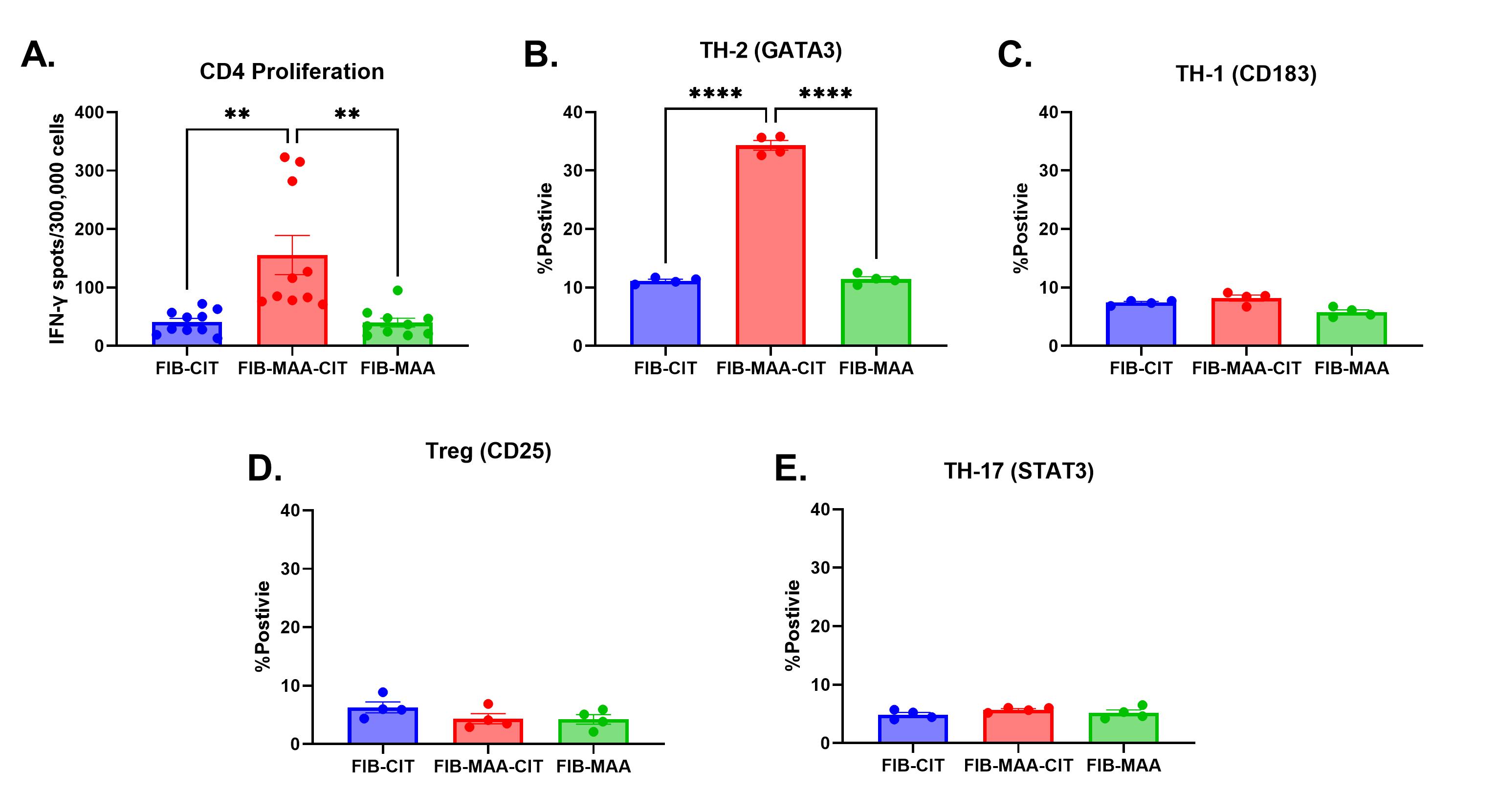
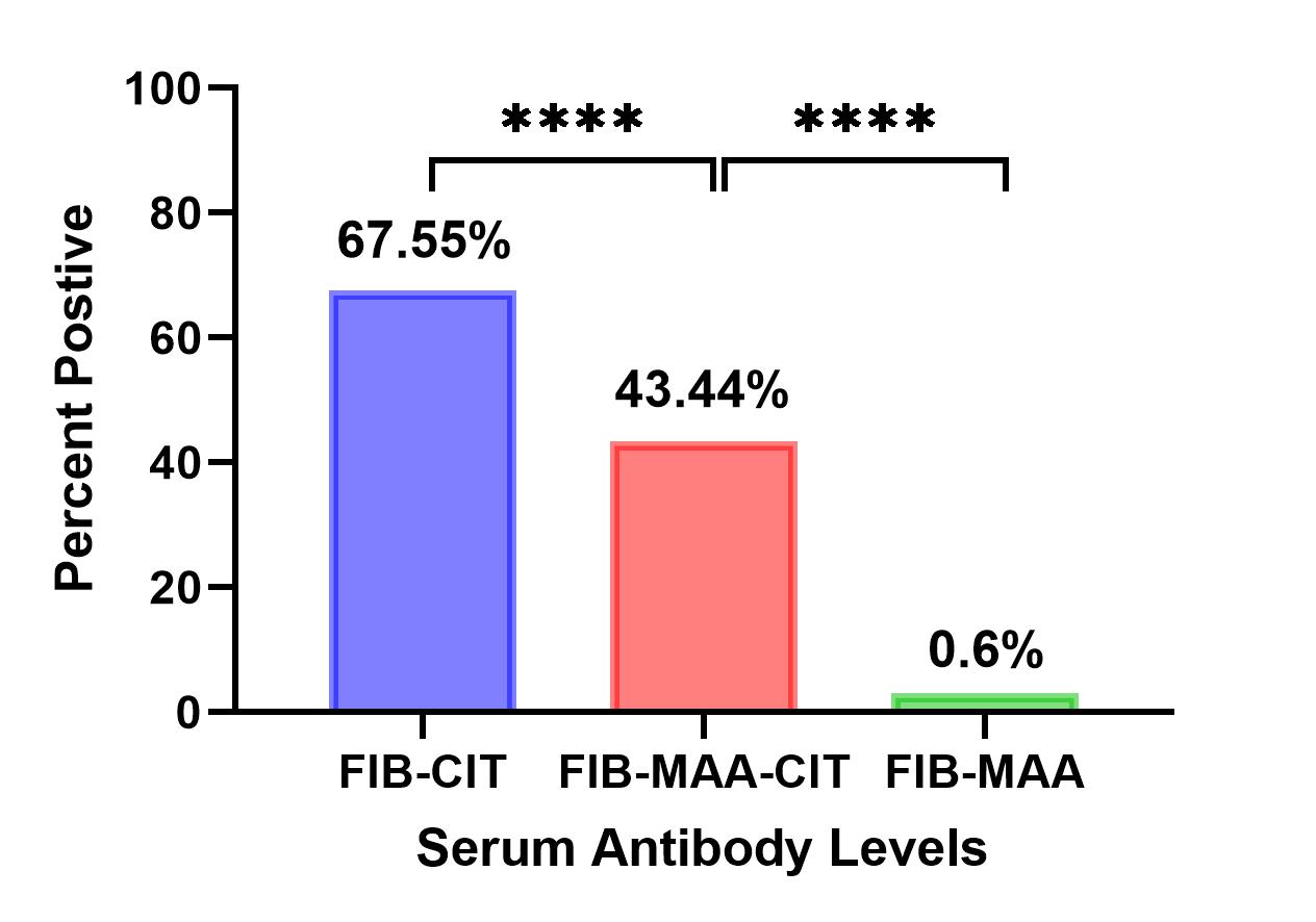
Results: Participant characteristics were similar in the 2 cohorts (90-91% male; mean age 63-73 years; 78-100% anti-CCP positive). CD4 cells proliferated significantly more to FIB-MAA-CIT than either FIB-CIT or FIB-MAA (p< 0.01) (Fig. 1A). This proliferation represented a predominant Th2 response based on increased GATA3 %-positivity in response to FIB-MAA-CIT vs. FIB-CIT or FIB-MAA (p< 0.0001) (Fig. 1B). In contrast, Th1 (CD183) (Fig. 1C), T-Reg (CD25) (Fig.1D) and Th17 (STAT3) %-positivity (Fig. 1E) showed no differences between FIB-CIT, FIB-MAA, and FIB-MAA-CIT. In VARA, the proportion of participants positive for serum IgG antibodies to FIB-CIT (68%) was significantly greater than positivity for either FIB-MAA-CIT (43%) or FIB-MAA (0.6%) (p˂0.0001) (Fig. 2).  

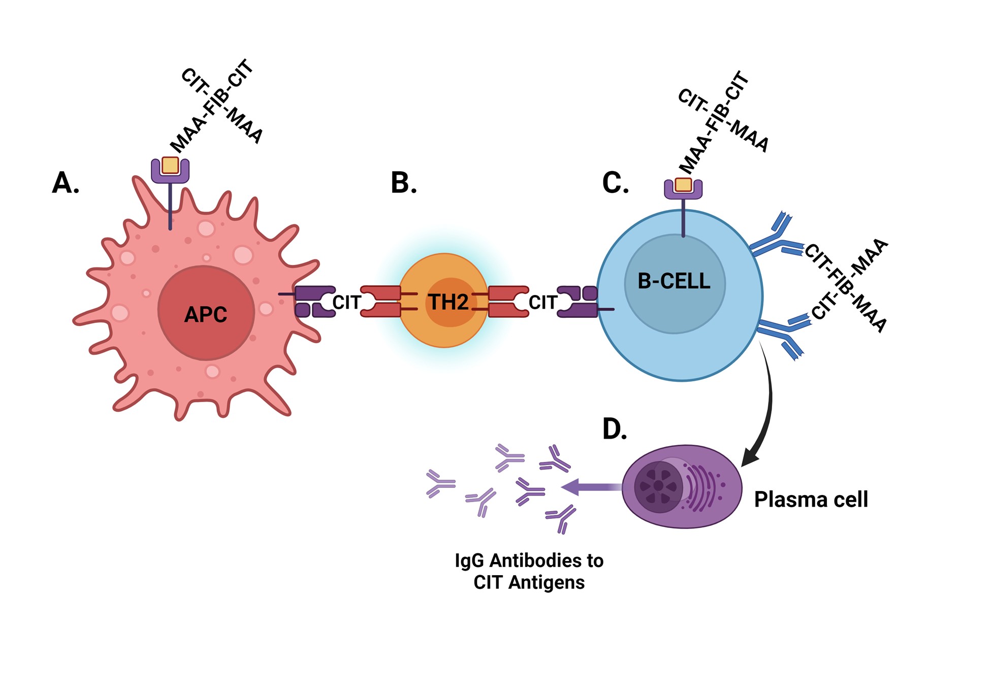
Conclusion: These results demonstrate that co-modification of CIT-protein with MAA significantly amplifies CD4 T cell proliferation and promotes a Th2 response to antigen stimulation. Given the known role of Th2 cells in promoting autoantibody expression, our data further suggest that resulting T cell responses promote the generation of ACPA but not anti-MAA antibody. Figure 3 provides a summary of the hypothesis for the mechanism behind these results.  Additional investigation is needed to understand whether these observations are driven by linked recognition (binding and uptake of co-modified antigen followed by preferential processing and presentation of CIT antigen) and whether interventions targeting MAA could reduce pathologic immune responses to CIT-protein in RA.

Supporting image 1

Figure 1. CD4 Proliferation and Percent Positivity of T Cell Phenotypes in Response to Modified Fibrinogen CD4 proliferation as a function of IFN-γ spots/300,000 cells (ELISpot); FIB-MAA-CIT significantly increased vs. FIB-CIT and FIB-MAA (**p<0.01) (A). %-positivity for Th2 (GATA3+) significantly increased (****p<0.0001) with FIB-MAA-CIT vs. FIB-CIT and FIB-MAA (B). Autologous T cells phenotyped via flow cytometry showed no differences in %-positivity for Th1 (CD183+) (C). No significant group difference in %-positivity for T-Reg (CD25+) (D) or Th17 (STAT3+) between cells stimulated with FIB-CIT, FIB-MAA, or FIB-MAA-CIT (E) (n=10).

Supporting image 2

Figure 2. Serum IgG Antibody Levels to Modified Fibrinogen. The proportion of study participants with RA positive for serum IgG antibodies to FIB-CIT was significantly increased compared to serum IgG antibodies to both FIB-MAA and FIB-MAA-CIT (****p˂0.0001) (N=2484)

Supporting image 3

Figure 3. Potential Mechanism by which Co-modification of a Protein with MAA and CIT Amplifies Both the T cell and Antibody Responses to CIT. MAA-CIT-modified protein initiates ACPA generation via MAA binding to scavenger receptors on Antigen Presenting Cells (APCs) (A). During the processing/presentation, CIT-modified peptides are presented via MHC Class II to T helper cells (Th; activated to Th2 phenotype) (B). B cells bind CIT or MAA-CIT via surface immunoglobulin and can process/present CIT-peptide in MHC Class II (C). With Th2 ‘help’, B cells differentiate to plasma cells and secrete ACPA (D).


Disclosures: B. Butler: None; W. Zhou: None; M. Duryee: None; N. Aripova: None; E. Sharp: None; C. Hunter: None; B. Kramer: None; H. Sayles: None; J. O'Dell: None; G. Thiele: None; B. England: Boehringer-Ingelheim, 5; J. Baker: Cumberland Pharma, 2, Formation Bio, 2, Horizon, 5; A. Reimold: None; G. Kerr: AstraZeneca, 1, Boehringer-Ingelheim, 6, Bristol-Myers Squibb(BMS), 1, CSL Behring, 6, Janssen, 6, Novartis, 1, Pfizer, 6, Sanofi, 6, UCB, 1; T. Mikuls: Elsevier, 9, Horizon Therapeutics, 2, 5, Pfizer, 2, Sanofi, 2, UCB Pharma, 2, Wolters Kluwer Health (UpToDate), 9.

To cite this abstract in AMA style:

Butler B, Zhou W, Duryee M, Aripova N, Sharp E, Hunter C, Kramer B, Sayles H, O'Dell J, Thiele G, England B, Baker J, Reimold A, Kerr G, Mikuls T. Co-modification of Citrullinated Proteins with Malondialdehyde-Acetaldehyde Leads to Amplified T Cell Responses and Increased Disease-specific Autoantibody Concentrations [abstract]. Arthritis Rheumatol. 2024; 76 (suppl 9). https://acrabstracts.org/abstract/co-modification-of-citrullinated-proteins-with-malondialdehyde-acetaldehyde-leads-to-amplified-t-cell-responses-and-increased-disease-specific-autoantibody-concentrations/. Accessed .
  • Tweet
  • Click to email a link to a friend (Opens in new window) Email
  • Click to print (Opens in new window) Print

« Back to ACR Convergence 2024

ACR Meeting Abstracts - https://acrabstracts.org/abstract/co-modification-of-citrullinated-proteins-with-malondialdehyde-acetaldehyde-leads-to-amplified-t-cell-responses-and-increased-disease-specific-autoantibody-concentrations/

Advanced Search

Your Favorites

You can save and print a list of your favorite abstracts during your browser session by clicking the “Favorite” button at the bottom of any abstract. View your favorites »

All abstracts accepted to ACR Convergence are under media embargo once the ACR has notified presenters of their abstract’s acceptance. They may be presented at other meetings or published as manuscripts after this time but should not be discussed in non-scholarly venues or outlets. The following embargo policies are strictly enforced by the ACR.

Accepted abstracts are made available to the public online in advance of the meeting and are published in a special online supplement of our scientific journal, Arthritis & Rheumatology. Information contained in those abstracts may not be released until the abstracts appear online. In an exception to the media embargo, academic institutions, private organizations, and companies with products whose value may be influenced by information contained in an abstract may issue a press release to coincide with the availability of an ACR abstract on the ACR website. However, the ACR continues to require that information that goes beyond that contained in the abstract (e.g., discussion of the abstract done as part of editorial news coverage) is under media embargo until 10:00 AM ET on November 14, 2024. Journalists with access to embargoed information cannot release articles or editorial news coverage before this time. Editorial news coverage is considered original articles/videos developed by employed journalists to report facts, commentary, and subject matter expert quotes in a narrative form using a variety of sources (e.g., research, announcements, press releases, events, etc.).

Violation of this policy may result in the abstract being withdrawn from the meeting and other measures deemed appropriate. Authors are responsible for notifying colleagues, institutions, communications firms, and all other stakeholders related to the development or promotion of the abstract about this policy. If you have questions about the ACR abstract embargo policy, please contact ACR abstracts staff at [email protected].

Wiley

  • Online Journal
  • Privacy Policy
  • Permissions Policies
  • Cookie Preferences

© Copyright 2025 American College of Rheumatology