ACR Meeting Abstracts

ACR Meeting Abstracts

  • Meetings
    • ACR Convergence 2024
    • ACR Convergence 2023
    • 2023 ACR/ARP PRSYM
    • ACR Convergence 2022
    • ACR Convergence 2021
    • ACR Convergence 2020
    • 2020 ACR/ARP PRSYM
    • 2019 ACR/ARP Annual Meeting
    • 2018-2009 Meetings
    • Download Abstracts
  • Keyword Index
  • Advanced Search
  • Your Favorites
    • Favorites
    • Login
    • View and print all favorites
    • Clear all your favorites
  • ACR Meetings

Abstract Number: 2521

Clinicopathologic Associations Between Macrophage Location and Functional Profile in Giant Cell Arteritis

Michael Putman1, Nader Khalidi2, Carol Langford3, Curry Koening4, Christian Pagnoux5, David Cuthbertson6, Carol McAlear7 and Peter Merkel7, and the Vasculitis Clinical Research Consortium (VCRC), 1The Medical College of Wisconsin, Milwaukee, WI, 2McMaster University, Hamilton, ON, Canada, 3Cleveland Clinic, Moreland Hills, OH, 4University of Texas Dell Medical School, Austin, TX, 5Mount Sinai Hospital, Toronto, ON, Canada, 6University of South Florida, Tampa, FL, 7University of Pennsylvania, Philadelphia, PA

Meeting: ACR Convergence 2024

Keywords: Epidemiology, giant cell arteritis, immunology, macrophages, Vasculitis

  • Tweet
  • Click to email a link to a friend (Opens in new window) Email
  • Click to print (Opens in new window) Print
Session Information

Date: Monday, November 18, 2024

Title: Vasculitis – Non-ANCA-Associated & Related Disorders Poster III

Session Type: Poster Session C

Session Time: 10:30AM-12:30PM

Background/Purpose: Macrophages play a central role in the pathogenesis of giant cell arteritis (GCA), but few studies have correlated macrophage phenotype or topography from temporal artery biopsy specimens with clinical outcomes. This project aimed to identify associations between the development of vascular damage and the location or functional profile of macrophages in temporal artery biopsy specimens.

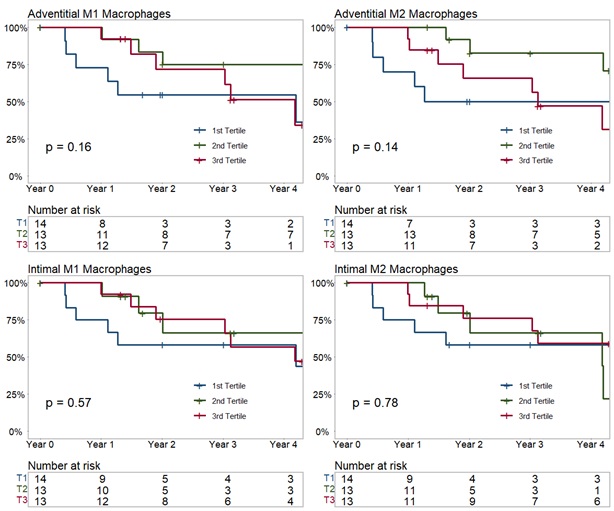
Methods: Patients from the Vasculitis Clinical Research Consortium (VCRC) Longitudinal Study of GCA with linked temporal artery biopsy specimens in the VCRC Tissue Repository were included. Temporal artery tissue was stained using immunofluorescent antibodies to CD3 (T Cells), CD4 (T cells), FoxP3 (T Regs), CD68/CD86 (inflammatory “M1” macrophages), and CD68/CD163/CD206 (remodeling “M2” macrophages) using an Opal 7-Color Automation IHC Kit. The relative proportions of these cell types as a percent of the total number of cells in the adventitia and intima were calculated. Associations between macrophage location (adventitia, intima) and phenotype (M1 or M2) and the Large-Vessel Vasculitis Index of Damage (LVVID) were evaluated using t testing and the Kaplan-Meier estimate.

Results: 40 patients had biopsy specimens available for analysis. The average age was 69.9 years (standard deviation [SD] 9.1), all patients were white, and 21/40 (52.5%) male. The mean LVVID score was 1.4 (SD 2.2) at baseline; a minority of patients developed progressive damage over the first year of observation (4/40, 11.4%, mean follow up time 314 days, SD 118 days). In unadjusted analysis there was no association between the developing new damage on the LVVID within the first year of observation and the relative abundance of intimal M1 macrophages (1.8% vs. 0.7% developed damage, p = 0.12), adventitial M1 macrophages (2.0% vs 1.2% developed damage, p = 0.53), intimal M2 macrophages (2.9% vs. 1.4% developed damage, p = 0.37), or adventitial M2 macrophages (3.7% vs. 2.4% developed damage, p = 0.61). There were also no associations identified for CD3+ or CD4+ T cells, but the relative abundance of adventitial FoxP3+ (TReg) cells was associated with developing damage (2.8% vs. 1.0% developed damage, p = 0.04). When the analysis period was extended from the first year of observation to include the entire observation period, half of patients developed damage (20/40, 50%, mean follow up time 1,109 days, SD 962 days. In unadjusted analysis over the entire observation period, there were no additional associations identified between developing damage and either macrophage location (intima or adventitia) or phenotype (M1 or M2). A time to event analysis using a Kaplan-Meier estimate supported these findings (Figure).

Conclusion: Macrophage topography and phenotype from temporal artery biopsy specimens are not associated with the development of damage in patients with GCA.

Supporting image 1

Figure 1: Estimate of time (days) to first damage on the Large-Vessel Vasculitis Index of Damage, stratified by macrophage location (adventitia or intima) and functional profile (“M1” (CD68/CD86+) or “M2” (CD68/163+ or CD68/CD206+)


Disclosures: M. Putman: AbbVie/Abbott, 5, Amgen, 5, AstraZeneca, 2, 5, GlaxoSmithKlein(GSK), 2, Novartis, 1, 5; N. Khalidi: AbbVie/Abbott, 5, Bristol-Myers Squibb(BMS), 5, GlaxoSmithKlein(GSK), 6, Otsuka, 1, Roche, 1; C. Langford: AbbVie/Abbott, 12, Non-paid consultant, AstraZeneca, 5, 12, Non-paid consultant, Bristol-Myers Squibb(BMS), 5, 12, Non-paid consultant, GlaxoSmithKlein(GSK), 5; C. Koening: Amgen, 6; C. Pagnoux: AstraZeneca, 2, 6, CSL Vifor, 2, GSK, 2, 6, Otsuka, 2, 6; D. Cuthbertson: None; C. McAlear: None; P. Merkel: AbbVie/Abbott, 2, 5, Amgen, 2, 5, argenx, 2, AstraZeneca, 2, 5, Boehringer Ingelheim, 3, 5, Bristol Myers Squibb, 2, 5, Cabaletta, 2, ChemoCentryx, 2, 5, CSL Behring, 2, Dynacure, 2, Eicos, 5, Electra, 5, EMDSerano, 2, Forbius, 2, 5, Genentech/Roche, 2, 5, Genzyme/Sanofi, 2, 5, GSK, 2, 5, HiBio, 2, Immagene, 2, InflaRx, 2, 5, Jannsen, 2, Kiniksa, 2, Kyverna, 2, Magenta, 2, MiroBio, 2, Neutrolis, 2, Novartis, 2, NS Pharma, 2, Pfizer, 2, Q32, 2, 11, Regeneron, 2, Sanofi, 5, Sparrow, 2, 11, Takeda, 2, 5, Talaris, 2, UpToDate, 9, Visterra, 2.

To cite this abstract in AMA style:

Putman M, Khalidi N, Langford C, Koening C, Pagnoux C, Cuthbertson D, McAlear C, Merkel P. Clinicopathologic Associations Between Macrophage Location and Functional Profile in Giant Cell Arteritis [abstract]. Arthritis Rheumatol. 2024; 76 (suppl 9). https://acrabstracts.org/abstract/clinicopathologic-associations-between-macrophage-location-and-functional-profile-in-giant-cell-arteritis/. Accessed .
  • Tweet
  • Click to email a link to a friend (Opens in new window) Email
  • Click to print (Opens in new window) Print

« Back to ACR Convergence 2024

ACR Meeting Abstracts - https://acrabstracts.org/abstract/clinicopathologic-associations-between-macrophage-location-and-functional-profile-in-giant-cell-arteritis/

Advanced Search

Your Favorites

You can save and print a list of your favorite abstracts during your browser session by clicking the “Favorite” button at the bottom of any abstract. View your favorites »

All abstracts accepted to ACR Convergence are under media embargo once the ACR has notified presenters of their abstract’s acceptance. They may be presented at other meetings or published as manuscripts after this time but should not be discussed in non-scholarly venues or outlets. The following embargo policies are strictly enforced by the ACR.

Accepted abstracts are made available to the public online in advance of the meeting and are published in a special online supplement of our scientific journal, Arthritis & Rheumatology. Information contained in those abstracts may not be released until the abstracts appear online. In an exception to the media embargo, academic institutions, private organizations, and companies with products whose value may be influenced by information contained in an abstract may issue a press release to coincide with the availability of an ACR abstract on the ACR website. However, the ACR continues to require that information that goes beyond that contained in the abstract (e.g., discussion of the abstract done as part of editorial news coverage) is under media embargo until 10:00 AM ET on November 14, 2024. Journalists with access to embargoed information cannot release articles or editorial news coverage before this time. Editorial news coverage is considered original articles/videos developed by employed journalists to report facts, commentary, and subject matter expert quotes in a narrative form using a variety of sources (e.g., research, announcements, press releases, events, etc.).

Violation of this policy may result in the abstract being withdrawn from the meeting and other measures deemed appropriate. Authors are responsible for notifying colleagues, institutions, communications firms, and all other stakeholders related to the development or promotion of the abstract about this policy. If you have questions about the ACR abstract embargo policy, please contact ACR abstracts staff at [email protected].

Wiley

  • Online Journal
  • Privacy Policy
  • Permissions Policies
  • Cookie Preferences

© Copyright 2025 American College of Rheumatology