ACR Meeting Abstracts

ACR Meeting Abstracts

  • Meetings
    • ACR Convergence 2024
    • ACR Convergence 2023
    • 2023 ACR/ARP PRSYM
    • ACR Convergence 2022
    • ACR Convergence 2021
    • ACR Convergence 2020
    • 2020 ACR/ARP PRSYM
    • 2019 ACR/ARP Annual Meeting
    • 2018-2009 Meetings
    • Download Abstracts
  • Keyword Index
  • Advanced Search
  • Your Favorites
    • Favorites
    • Login
    • View and print all favorites
    • Clear all your favorites
  • ACR Meetings

Abstract Number: 1763

Circulating NK and CD8+ Cytotoxic T Cells in Treatment Naïve JDM Demonstrate Higher Cytotoxic and Interferon Signature as Compared to Childhood-Onset SLE

Qinmengge Li1, Celine Berthier1, Christine Goudsmit1, Sophia Matossian1, Rachael Wasikowski2, Johann Gudjonsson1, J. Michelle Kahlenberg1, Lam C. Tsoi2 and Jessica Turnier3, 1University of Michigan, Ann Arbor, MI, 2Michigan, Dept. of Dermatology, Ann Arbor, MI, 3University of Michigan, Saline, MI

Meeting: ACR Convergence 2024

Keywords: Bioinformatics, Myositis, Natural Killer Cells, Pediatric rheumatology, Systemic lupus erythematosus (SLE)

  • Tweet
  • Click to email a link to a friend (Opens in new window) Email
  • Click to print (Opens in new window) Print
Session Information

Date: Monday, November 18, 2024

Title: Pediatric Rheumatology – Basic Science Poster

Session Type: Poster Session C

Session Time: 10:30AM-12:30PM

Background/Purpose: Juvenile dermatomyositis (JDM) and childhood-onset systemic lupus erythematosus (cSLE) are pediatric autoimmune diseases that can present with overlapping clinical features yet have unique tropism for major organ involvement. The objective of this study was to characterize differences in immune cell proportions and transcriptional signatures in treatment naïve (TN) JDM vs. cSLE to identify disease-specific mechanisms and treatment targets.

Methods: We isolated fresh peripheral blood mononuclear cells (PBMCs) from TN JDM (n=5), TN cSLE (n=4) and healthy controls (CTL; n=4). All patients met 2017 EULAR/ACR classification criteria for JDM or 2019 ACR/EULAR classification criteria for SLE, respectively. Single cell RNA sequencing was performed on the 10x Genomics platform. Ambient RNA and doublets were removed by SoupX and scDblFinder, respectively. We performed batch correction using Harmony, dimensionality reduction using principal component analysis and uniform manifold approximation and projection, and clustering using shared nearest neighbor. Cell types were annotated via published cell type specific marker genes. We analyzed (1) relative abundance of each cell type within disease and control by testing pair-wise significance using z-test and (2) differential transcriptomic signatures per cell type between JDM and cSLE by using likelihood ratio test and reported log fold change (logFC) and Bonferroni corrected p-values.

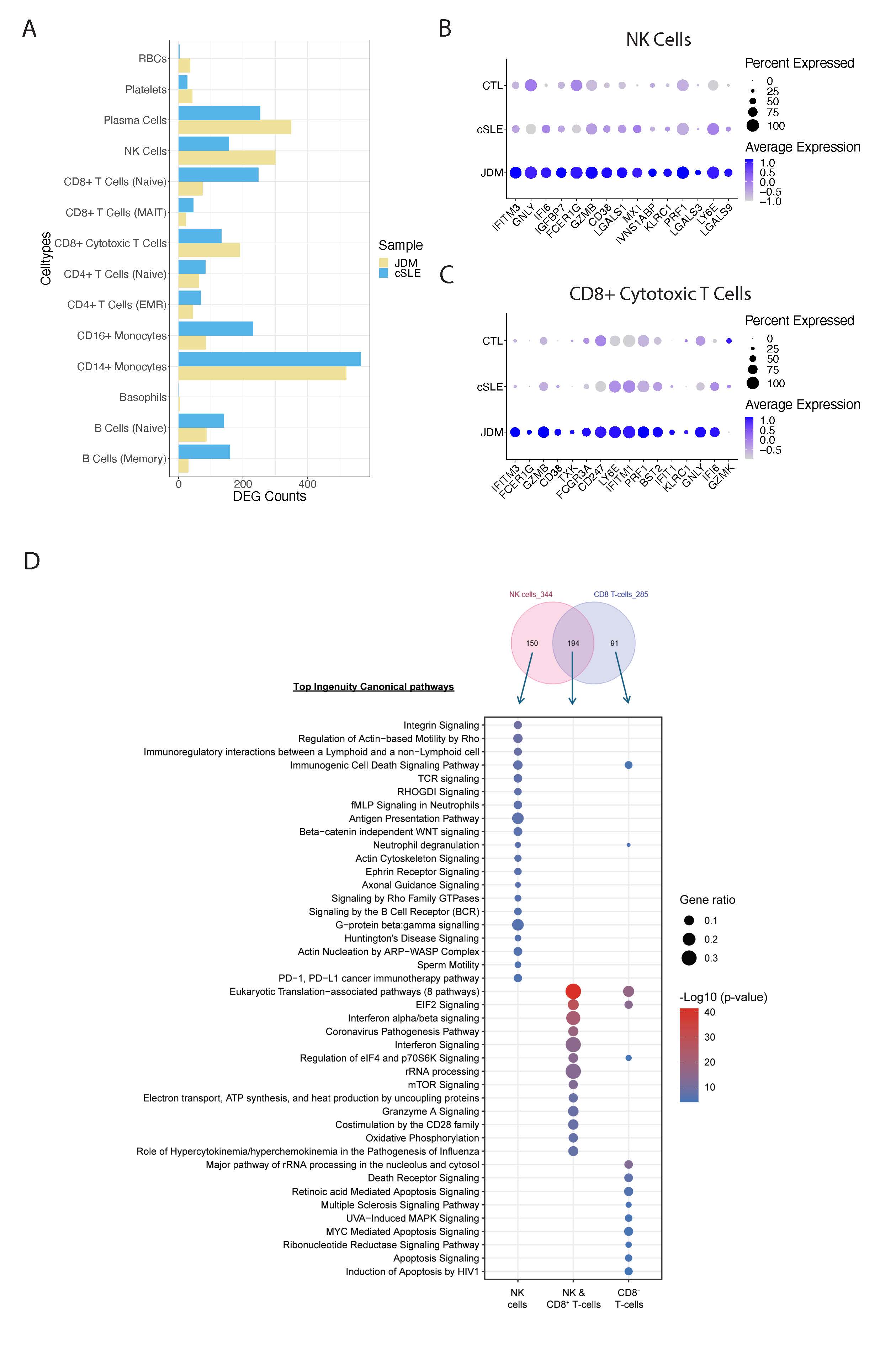
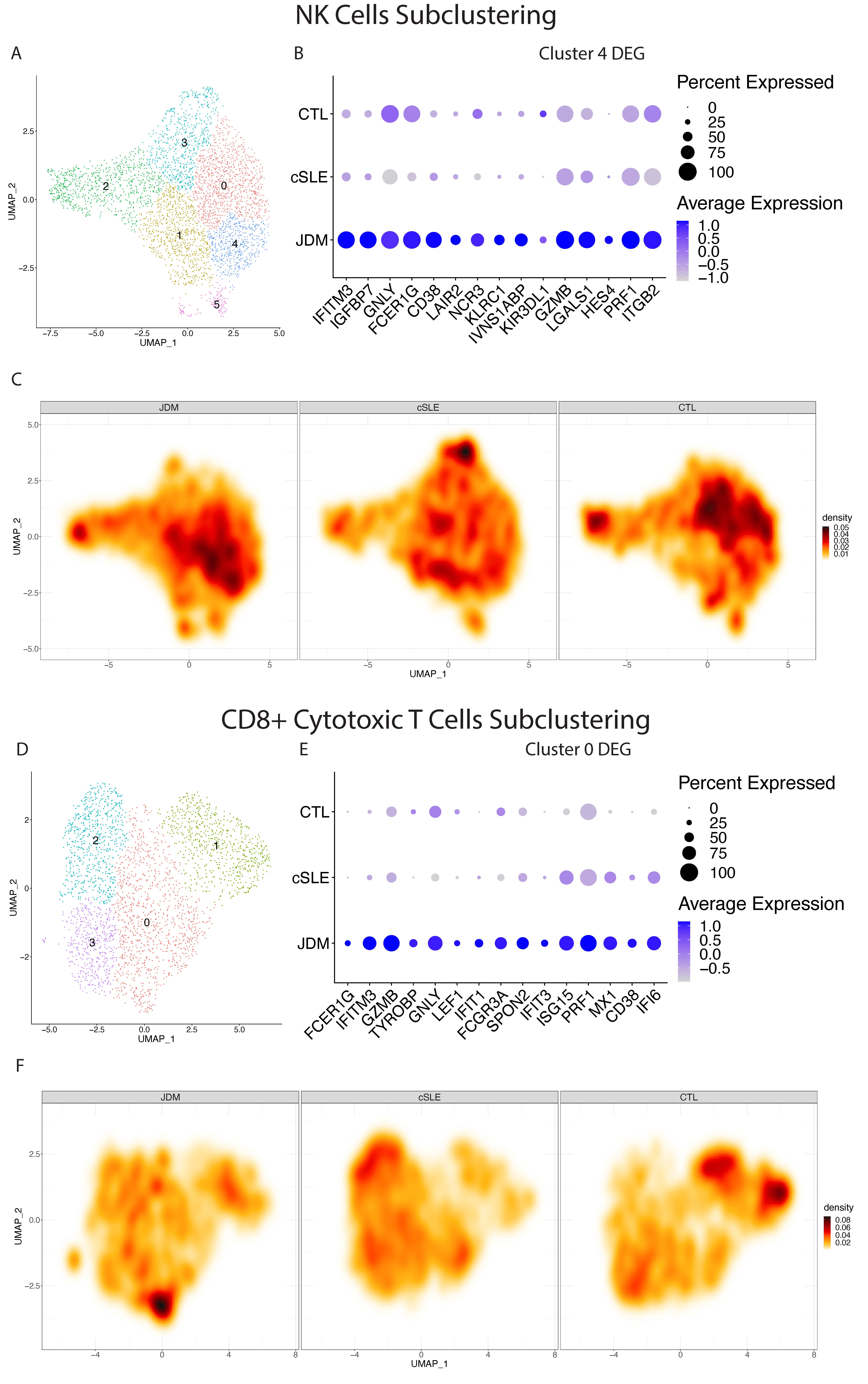
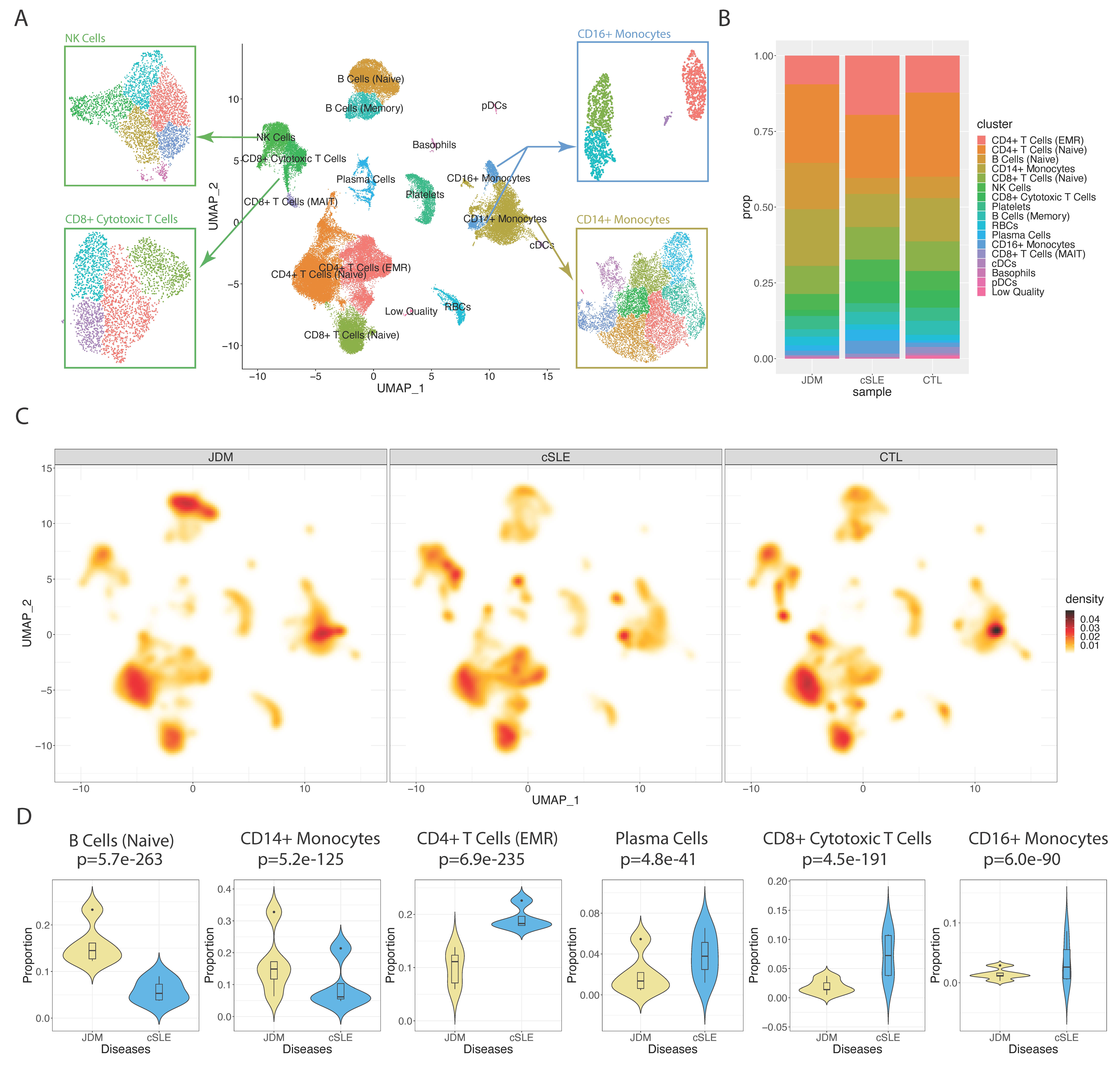
Results: 16 distinct annotated cell types were identified from 67,661 cells (Fig 1A). As compared to cSLE, JDM demonstrated increased relative abundance of naïve B-cells and CD14+ monocytes and decreased relative abundance of CD4+ effector memory and regulatory (EMR) T-cells, plasma cells, CD8+ cytotoxic T-cells and CD16+ monocytes (Fig 1B-D). Cytotoxic cells in JDM, including NK cells and CD8+ cytotoxic T cells, displayed a higher number of DE genes (DEG) as compared to cSLE (Fig 2A). JDM cytotoxic cells displayed a common increased cytotoxic and IFN signature, with 194 shared DEG, including GNLY, IFITM3, FCER1G, GZMB and CD38 in the top 10 DEG (Fig 2B-D). Interestingly, JDM CD8+ cytotoxic T-cells had increased GZMB expression as compared to increased GZMK in cSLE (Fig 2C). NK cells in JDM were uniquely enriched for antigen presentation, PD-L1 signaling and fMLP signaling in neutrophils, and CD8+ cytotoxic cells demonstrated enrichment for death receptor and apoptosis signaling (Fig 2D). Subclustering of cytotoxic cells highlighted an increase in NK subcluster 4 in JDM (Fig 3A & 3C), which had higher expression of immunoregulatory receptors of cytotoxicity, including NCR3, KLRC1 and KIR3DL1 (Fig 3B). JDM also had more cells in CD8+ cytotoxic T-cell subcluster 0 (Fig 3D & 3F), which was notable for increased expression of TYROBP, an adaptor protein mediating activation of multiple immune cell types (Fig 3E).

Conclusion: We identified differences in immune cell proportions in TN JDM vs. cSLE. We highlight cytotoxic cells in JDM vs. cSLE as displaying higher IFN, apoptotic and chemotactic signaling. Further investigation between JDM and cSLE-specific single-cell transcriptomic signatures has the potential to identify more specific treatments and biomarkers for each disease.

Supporting image 1

Figure 1. Cell types identified and relative cell type abundance in TN JDM vs. cSLE. A, Uniform manifold approximation and projection (UMAP) visualization of cell type annotation with highlights of NK cells, CD8+ cytotoxic T cells, CD16+ monocytes and CD14+ monocytes sub-clustering. B, Cell type proportion bar plot comparing disease and control samples. C, UMAP density plot showing the cell density over UMAP grid comparing disease and control samples. D, Violin plot comparing cell type proportions between TN JDM and cSLE; P-values are derived from z-test.

Supporting image 2

Figure 2. Differential transcriptomic signatures per cell type between JDM and cSLE. A, Bar plot showing the number of up-regulated genes comparing JDM and cSLE in each cell type. B, Dot plot of top up-regulated genes of JDM in NK cells. C, Dot plot of top up-regulated genes of JDM and GZMK in CD8+ cytotoxic T cells. D, Comparison of the NK and CD8+ T-cell genes: the top panel displays the Venn diagram of DEGs numbers in common between and unique to each NK cells and CD8+ cytotoxic T cells; the bottom panel displays the top 20 ingenuity canonical pathways (p-value<0.05) from each gene list.

Supporting image 3

Figure 3. Cytotoxic immune cell sub-clustering results. A, UMAP visualization of clustered groups in NK cells. B, Dot plot of top up-regulated genes of JDM in NK cells sub-cluster 4. C, UMAP density plot showing the cell density over UMAP grid comparing disease and control samples for NK cells. D, UMAP visualization of clustered groups in CD8+ cytotoxic T cells. E, Dot plot of top up-regulated genes of JDM in CD8+ cytotoxic T cells sub-cluster 0. F, UMAP density plot showing the cell density over UMAP grid comparing disease and control samples for CD8+ cytotoxic T cells.


Disclosures: Q. Li: None; C. Berthier: None; C. Goudsmit: None; S. Matossian: None; R. Wasikowski: None; J. Gudjonsson: AbbVie/Abbott, 12, support, Boehringer-Ingelheim, 12, support, Bristol-Myers Squibb(BMS), 12, support, Eli Lilly, 12, support, Janssen, 12, support, Novartis, 12, support; J. Kahlenberg: Amgen, 12, coauthor on publication, AstraZeneca, 2, Bristol-Myers Squibb(BMS), 2, 5, Eli Lilly, 2, Gilead, 2, GlaxoSmithKlein(GSK), 2, Janssen, 5; L. Tsoi: Janssen, 5; J. Turnier: Cabaletta Bio, 1.

To cite this abstract in AMA style:

Li Q, Berthier C, Goudsmit C, Matossian S, Wasikowski R, Gudjonsson J, Kahlenberg J, Tsoi L, Turnier J. Circulating NK and CD8+ Cytotoxic T Cells in Treatment Naïve JDM Demonstrate Higher Cytotoxic and Interferon Signature as Compared to Childhood-Onset SLE [abstract]. Arthritis Rheumatol. 2024; 76 (suppl 9). https://acrabstracts.org/abstract/circulating-nk-and-cd8-cytotoxic-t-cells-in-treatment-naive-jdm-demonstrate-higher-cytotoxic-and-interferon-signature-as-compared-to-childhood-onset-sle/. Accessed .
  • Tweet
  • Click to email a link to a friend (Opens in new window) Email
  • Click to print (Opens in new window) Print

« Back to ACR Convergence 2024

ACR Meeting Abstracts - https://acrabstracts.org/abstract/circulating-nk-and-cd8-cytotoxic-t-cells-in-treatment-naive-jdm-demonstrate-higher-cytotoxic-and-interferon-signature-as-compared-to-childhood-onset-sle/

Advanced Search

Your Favorites

You can save and print a list of your favorite abstracts during your browser session by clicking the “Favorite” button at the bottom of any abstract. View your favorites »

All abstracts accepted to ACR Convergence are under media embargo once the ACR has notified presenters of their abstract’s acceptance. They may be presented at other meetings or published as manuscripts after this time but should not be discussed in non-scholarly venues or outlets. The following embargo policies are strictly enforced by the ACR.

Accepted abstracts are made available to the public online in advance of the meeting and are published in a special online supplement of our scientific journal, Arthritis & Rheumatology. Information contained in those abstracts may not be released until the abstracts appear online. In an exception to the media embargo, academic institutions, private organizations, and companies with products whose value may be influenced by information contained in an abstract may issue a press release to coincide with the availability of an ACR abstract on the ACR website. However, the ACR continues to require that information that goes beyond that contained in the abstract (e.g., discussion of the abstract done as part of editorial news coverage) is under media embargo until 10:00 AM ET on November 14, 2024. Journalists with access to embargoed information cannot release articles or editorial news coverage before this time. Editorial news coverage is considered original articles/videos developed by employed journalists to report facts, commentary, and subject matter expert quotes in a narrative form using a variety of sources (e.g., research, announcements, press releases, events, etc.).

Violation of this policy may result in the abstract being withdrawn from the meeting and other measures deemed appropriate. Authors are responsible for notifying colleagues, institutions, communications firms, and all other stakeholders related to the development or promotion of the abstract about this policy. If you have questions about the ACR abstract embargo policy, please contact ACR abstracts staff at [email protected].

Wiley

  • Online Journal
  • Privacy Policy
  • Permissions Policies
  • Cookie Preferences

© Copyright 2025 American College of Rheumatology