ACR Meeting Abstracts

ACR Meeting Abstracts

  • Meetings
    • ACR Convergence 2024
    • ACR Convergence 2023
    • 2023 ACR/ARP PRSYM
    • ACR Convergence 2022
    • ACR Convergence 2021
    • ACR Convergence 2020
    • 2020 ACR/ARP PRSYM
    • 2019 ACR/ARP Annual Meeting
    • 2018-2009 Meetings
    • Download Abstracts
  • Keyword Index
  • Advanced Search
  • Your Favorites
    • Favorites
    • Login
    • View and print all favorites
    • Clear all your favorites
  • ACR Meetings

Abstract Number: 2540

Characterizing the Functional Role of Type I Interferons in Inflammatory Arthritis

Mary Huang, Richard Bell, Toolika Singh and Lionel Ivashkiv, Hospital for Special Surgery, New York, NY

Meeting: ACR Convergence 2024

Keywords: innate immunity, interferon, Mouse Models, RA, rheumatoid arthritis

  • Tweet
  • Email
  • Print
Session Information

Date: Monday, November 18, 2024

Title: Abstracts: RA – Animal Models

Session Type: Abstract Session

Session Time: 1:00PM-2:30PM

Background/Purpose: Rheumatoid arthritis (RA) is a chronic autoimmune disease characterized by synovial inflammation due to cellular infiltration and inflammatory mediators. Type I interferons (IFN) have been implicated in the pathogenesis and modulation of autoimmune diseases, including RA, but their role in RA pathogenesis remains poorly understood, partly owing to their complex association with immunity and tolerance. We aim to understand the role of type I IFN signaling in arthritis.

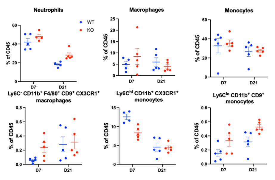
Methods: Zymosan-induced arthritis (ZIA) was induced in IFN alpha receptor 1 null mice (IFNAR1 KO) and C57BL/6J wild-type controls with a 180 µg zymosan intra-articular injection in the right knee using a protocol approved by the Weill Cornell Medicine IACUC. Ipsilateral and contralateral joints were dissected on D7 en bloc for downstream micro-computed tomography (µCT) and histologic analysis to assess synovitis and bone erosion (n=4/group). Slides were H&E-stained and analyzed via automated histomorphometry for synovial area and cell count. The proximal femurs were dissected and the bone marrow was flushed and analyzed for hematopoietic stem and progenitor cells (HSPCs) via flow cytometry (n=4/group). On D7 and D21, peri-articular synovial tissue of the ipsilateral joint was collected for flow cytometry analysis at D7 and D21 (n=5/group). Appropriate t-test and 2-way ANOVA statistical tests were perfomed. 

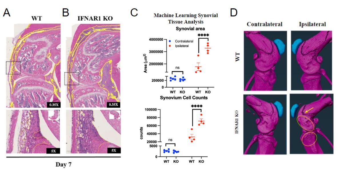
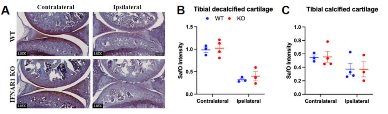
Results: Histologic and µCT analysis of the ipsilateral joint from D7 males revealed significantly increased synovial area (Fig 1 WT: 1.7 x 106 μm2 ± 6.3 x 105 μm2 vs IFNAR KO: 3.2 x 106 μm2 ± 3.7 x 105 μm2, p< 0.01), synovial cell count (WT: 3.2 x 10⁴ cells ± 1.3×10⁴ cells vs KO: 7.1 x 10⁴ cells ± 1.1 x 10⁴ cells, p< 0.01) and bone erosion in IFNAR1 KO mice compared to WT on D7 post zymosan injection (Fig 1). We did not observe changes in levels of Lin-Sca1+cKit+ HSPCs, an early progenitor cell, multipotent progenitors (MPPs), short-term hematopoietic stem cells (ST-HSCs), long-term HSCs (LT-HSCs), or MPP2, MPP3, and MPP4 cells in KO with ZIA (not shown). Tibial cartilage proteoglycan content did not differ between KO and WT (Fig 2). Flow cytometry analysis of synovial tissue from the ipsilateral knee shows significantly increased neutrophils at D21 (p=0.05) and significantly decreased Ly6ChiCD11b+CX3CR1+ monocytes at D7 (p=0.01) (Fig 3). 

Conclusion: Our data demonstrate that ZIA in IFNAR1 KO increases synovitis at D7, but does not affect myelopoiesis or cartilage degradation at day 7 post zymosan injection. Unexpectedly, levels of LSK cells and MPPs were unchanged at D7 in IFNAR1 KO despite increased synovitis. However, defining potential epigenetic and transcriptomic changes in bone marrow HSPCs and myeloid progenitors in IFNAR1 KO subjected to ZIA remains an area of future investigation. The observed increased inflammatory neutrophils yet decreased Ly6ChiCD11b+CX3CR1+ monocytes, which may be an inflammatory monocyte population, recapitulates the functionally heterogeneous outcomes known to occur downstream of IFNAR1. Further characterization of IFNAR1 KO may help resolve the cell-type specific activities catalyzed by IFNAR1 and has the potential to enable future type I interferon-specific therapeutics for inflammatory disease. 

Supporting image 1

Fig 1. Histological and micro-computed tomography (μCT) analysis at day 7 post intra-articular zymosan injection displays increased synovitis, greater synovial area and cell counts, and qualitatively increased porosity and bone erosion in IFNAR KO. Representative H&E stained slides of ipsilateral joints from WT (A) and IFNAR KO (B) mice with low (top panels) and high (bottom panels) magnification. Synovial area (yellow outline in A & B) and synovial cell counts (n = 4 per group) were assessed using an automated multi-scale computational pathotyping analysis (C). Representative μCT from n = 4 per group (D).

Supporting image 2

Fig 2. Zymosan-induced arthritis induces tibial cartilage proteoglycan loss in IFNAR1 KO and WT mice. (A) SafO stained slides of ipsilateral and contralateral joints from WT and IFNAR1 KO (n = 3-4 each group). SafO intensity of a 50,000 micrometer squared minimum annotation of tibial decalcified (B) or calcified cartilage (C) in the central region of the tibial plateau was quantified using QuPath.

Supporting image 3

Fig 3. IFNAR KO increases neutrophils at day 21 and decreases Ly6C-CD11b+F4/80+CD9+CX3CR1+ macrophages at day 7. Flow cytometry of peri-articular synovial tissue from the ipsilateral knee (n= 5 per group). Neutrophils, CD45+LyC+Ly6G+; Macrophages, Ly6C-CD11b+F4/80+; monocytes, Ly6C+CD11b+F4/80+.


Disclosures: M. Huang: None; R. Bell: None; T. Singh: None; L. Ivashkiv: None.

To cite this abstract in AMA style:

Huang M, Bell R, Singh T, Ivashkiv L. Characterizing the Functional Role of Type I Interferons in Inflammatory Arthritis [abstract]. Arthritis Rheumatol. 2024; 76 (suppl 9). https://acrabstracts.org/abstract/characterizing-the-functional-role-of-type-i-interferons-in-inflammatory-arthritis/. Accessed .
  • Tweet
  • Email
  • Print

« Back to ACR Convergence 2024

ACR Meeting Abstracts - https://acrabstracts.org/abstract/characterizing-the-functional-role-of-type-i-interferons-in-inflammatory-arthritis/

Advanced Search

Your Favorites

You can save and print a list of your favorite abstracts during your browser session by clicking the “Favorite” button at the bottom of any abstract. View your favorites »

All abstracts accepted to ACR Convergence are under media embargo once the ACR has notified presenters of their abstract’s acceptance. They may be presented at other meetings or published as manuscripts after this time but should not be discussed in non-scholarly venues or outlets. The following embargo policies are strictly enforced by the ACR.

Accepted abstracts are made available to the public online in advance of the meeting and are published in a special online supplement of our scientific journal, Arthritis & Rheumatology. Information contained in those abstracts may not be released until the abstracts appear online. In an exception to the media embargo, academic institutions, private organizations, and companies with products whose value may be influenced by information contained in an abstract may issue a press release to coincide with the availability of an ACR abstract on the ACR website. However, the ACR continues to require that information that goes beyond that contained in the abstract (e.g., discussion of the abstract done as part of editorial news coverage) is under media embargo until 10:00 AM ET on November 14, 2024. Journalists with access to embargoed information cannot release articles or editorial news coverage before this time. Editorial news coverage is considered original articles/videos developed by employed journalists to report facts, commentary, and subject matter expert quotes in a narrative form using a variety of sources (e.g., research, announcements, press releases, events, etc.).

Violation of this policy may result in the abstract being withdrawn from the meeting and other measures deemed appropriate. Authors are responsible for notifying colleagues, institutions, communications firms, and all other stakeholders related to the development or promotion of the abstract about this policy. If you have questions about the ACR abstract embargo policy, please contact ACR abstracts staff at [email protected].

Wiley

  • Online Journal
  • Privacy Policy
  • Permissions Policies
  • Cookie Preferences

© Copyright 2025 American College of Rheumatology