ACR Meeting Abstracts

ACR Meeting Abstracts

  • Meetings
    • ACR Convergence 2024
    • ACR Convergence 2023
    • 2023 ACR/ARP PRSYM
    • ACR Convergence 2022
    • ACR Convergence 2021
    • ACR Convergence 2020
    • 2020 ACR/ARP PRSYM
    • 2019 ACR/ARP Annual Meeting
    • 2018-2009 Meetings
    • Download Abstracts
  • Keyword Index
  • Advanced Search
  • Your Favorites
    • Favorites
    • Login
    • View and print all favorites
    • Clear all your favorites
  • ACR Meetings

Abstract Number: 1848

Characterizing Nailfold Capillary Changes in Dermatomyositis with a Dermatoscope

Josh Dan1, Grant Sprow2, Josef Concha3, Nilesh Kodali4, DeAnna Diaz5, Felix Chin6, Thomas Vazquez7 and Victoria Werth3, 1Philadelphia VAMC, Philadelphia, PA, USA and Department of Dermatology, Perelman School of Medicine at the University of Pennsylvania, Philadelphia, 2Albert Einstein College of Medicine, Philadelphia, PA, 3University of Pennsylvania, Philadelphia, PA, 4New Jersey Medical School, Coppell, TX, 5Philadelphia College of Medicine, Philadelphia, PA, 6University of Pennsylvania Perelman School of Medicine, Philadelphia, PA, 7FIU Wertheim College of Medicine, Virginia Beach, VA

Meeting: ACR Convergence 2022

Keywords: autoimmune diseases, Cutaneous, Dermatology, dermatomyositis, skin

  • Tweet
  • Email
  • Print
Session Information

Date: Monday, November 14, 2022

Title: Miscellaneous Rheumatic and Inflammatory Diseases Poster III

Session Type: Poster Session D

Session Time: 1:00PM-3:00PM

Background/Purpose: Nailfold capillary (NC) abnormalities are increasingly utilized in the evaluation of rheumatic conditions. Their presence can distinguish primary Raynaud’s phenomenon from secondary etiologies and are used in the diagnostic criteria of scleroderma. Dermoscopy is a convenient method of evaluating NC changes with similar efficacy to capillaroscopy. Literature pertaining to nailfold capillary changes in dermatomyositis (DM) commonly utilizes nailfold videocapillaroscopy to assess capillaries (200x magnification), which is not practical for clinical use. We aim to characterize nailfold capillary changes exhibited in dermatomyositis using a dermatoscope.

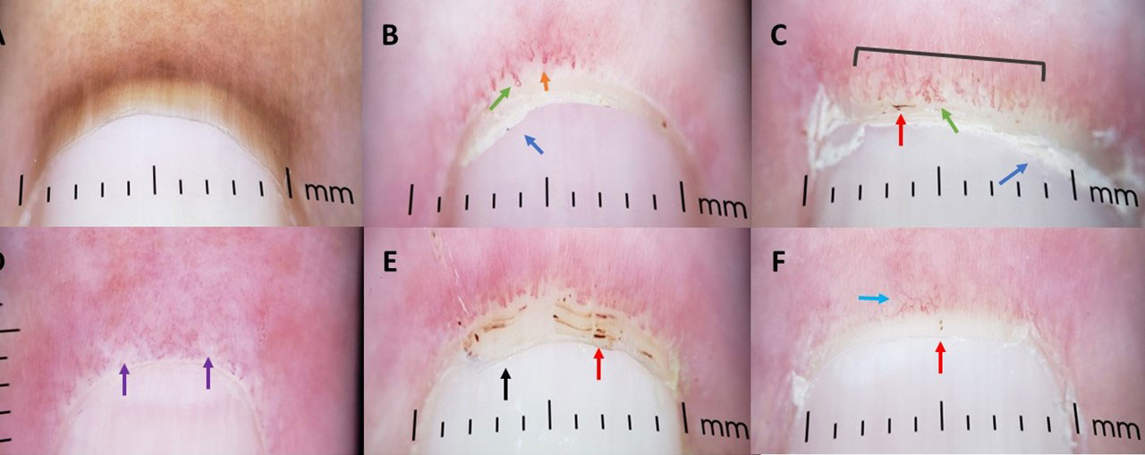
Methods: We imaged all ten nailfolds of subjects with dermatomyositis (DM) using a dermatoscope with a smartphone adapter. Fingernails were cleaned with an alcohol swab prior to evaluation. Subjects were included in they met ACR/EULAR criteria for DM or at the discretion of the principal investigator (VPW). Subjects with hypertension, hyperlipidemia, diabetes, glaucoma, or other primary rheumatic disease were excluded. Images were assessed for loop dilation, abnormal morphology, capillary bed disorganization, capillary hemorrhage, capillary dropout, decreased capillary density, subpapillary plexus visibility, cuticular dystrophy, and cuticular overgrowth (Figure 1). Cutaneous Dermatomyositis Area and Severity activity (CDASI-A) and damage (CDASI-D) scores were collected. Analysis used Student’s t-test and Chi-Squared test.

Results: 44 subjects, 17 with amyopathic DM (ADM), were included. Mean (standard error) CDASI activity and damage scores were 13 (1.05) and 4 (0.39), respectively. 89% of subjects demonstrated capillary abnormalities. Loop dilation (77%), followed by capillary bed disorganization (73%), were the two most common findings. NC changes were most often found on 4th and 5th fingers. Thumbs demonstrated the fewest abnormalities. Mean CDASI-A was higher in patients with abnormal morphology (CDASI 16 vs 10, p=0.003), capillary dropout (15 vs 10, p=0.016), decreased capillary density (17 vs 10, p< 0.001), and trended towards significance for loop dilation and capillary bed disorganization. There was a trend towards a higher percentage of capillary hemorrhage in patients with ADM compared to those with muscle disease (no difference in CDASI-A). Subjects with skin limited disease (n=23) vs active systemic disease demonstrated more abnormal morphology (74% vs 38%, p=0.02; no difference in CDASI-A).

Conclusion: Given their prevalence, dermatoscopic evaluation of nailfold capillary changes may be useful for the diagnosis of DM. Loop dilation and capillary bed disorganization are the two most common nailfold capillary changes in DM. Changes are most commonly found on the 4th and 5th digits. Abnormal morphology, capillary dropout, and decreased capillary correlate with cutaneous disease activity.

Supporting image 1

Figure 1: A. Normal nailfold capillary B. Abnormal morphology (green), dilated capillary loop (orange), cuticular dystrophy (blue) C. Capillary hemorrhage (red), abnormal morphology with dilation (green), cuticular dystrophy (blue), and overall disorganization of the capillary bed. D. Two areas of capillary dropout (purple) E. Cuticular overgrowth (black) and confluent areas of hemorrhage (red) F. Subpapillary plexus visibility (light blue) and two punctate hemorrhages (red).


Disclosures: J. Dan, None; G. Sprow, None; J. Concha, None; N. Kodali, None; D. Diaz, None; F. Chin, None; T. Vazquez, None; V. Werth, None.

To cite this abstract in AMA style:

Dan J, Sprow G, Concha J, Kodali N, Diaz D, Chin F, Vazquez T, Werth V. Characterizing Nailfold Capillary Changes in Dermatomyositis with a Dermatoscope [abstract]. Arthritis Rheumatol. 2022; 74 (suppl 9). https://acrabstracts.org/abstract/characterizing-nailfold-capillary-changes-in-dermatomyositis-with-a-dermatoscope/. Accessed .
  • Tweet
  • Email
  • Print

« Back to ACR Convergence 2022

ACR Meeting Abstracts - https://acrabstracts.org/abstract/characterizing-nailfold-capillary-changes-in-dermatomyositis-with-a-dermatoscope/

Advanced Search

Your Favorites

You can save and print a list of your favorite abstracts during your browser session by clicking the “Favorite” button at the bottom of any abstract. View your favorites »

All abstracts accepted to ACR Convergence are under media embargo once the ACR has notified presenters of their abstract’s acceptance. They may be presented at other meetings or published as manuscripts after this time but should not be discussed in non-scholarly venues or outlets. The following embargo policies are strictly enforced by the ACR.

Accepted abstracts are made available to the public online in advance of the meeting and are published in a special online supplement of our scientific journal, Arthritis & Rheumatology. Information contained in those abstracts may not be released until the abstracts appear online. In an exception to the media embargo, academic institutions, private organizations, and companies with products whose value may be influenced by information contained in an abstract may issue a press release to coincide with the availability of an ACR abstract on the ACR website. However, the ACR continues to require that information that goes beyond that contained in the abstract (e.g., discussion of the abstract done as part of editorial news coverage) is under media embargo until 10:00 AM ET on November 14, 2024. Journalists with access to embargoed information cannot release articles or editorial news coverage before this time. Editorial news coverage is considered original articles/videos developed by employed journalists to report facts, commentary, and subject matter expert quotes in a narrative form using a variety of sources (e.g., research, announcements, press releases, events, etc.).

Violation of this policy may result in the abstract being withdrawn from the meeting and other measures deemed appropriate. Authors are responsible for notifying colleagues, institutions, communications firms, and all other stakeholders related to the development or promotion of the abstract about this policy. If you have questions about the ACR abstract embargo policy, please contact ACR abstracts staff at [email protected].

Wiley

  • Online Journal
  • Privacy Policy
  • Permissions Policies
  • Cookie Preferences

© Copyright 2025 American College of Rheumatology