ACR Meeting Abstracts

ACR Meeting Abstracts

  • Meeting Abstracts
    • All Meetings
    • PRSYM 2026
    • ACR Convergence 2025
    • Download Abstract Supplements
  • Keyword Index
  • Search
  • My Favorites
    • View and print all favorites
    • Clear all favorites

Abstract Number: 1797

Characterizing Memory T Cell Subsets Associated with SLE Etiopathogenesis

Carol Nassar1, Rene Quevedo2, M. Teresa Ciudad2, Zoha Faheem3, Kieran Manion4, Carolina Munoz-Grajales5, Michael Kim6, Dafna Gladman7, Murray Urowitz8, Zahi Touma1, Tracy McGaha9 and Joan Wither6, 1University of Toronto, Toronto, ON, Canada, 2Princess Margaret Cancer Centre, Toronto, ON, Canada, 3UHN, Toronto, ON, Canada, 4Toronto Western Hospital, Toronto, ON, Canada, 5UHN/TWH, Toronto, ON, Canada, 6University Health Network, Toronto, ON, Canada, 7University of Toronto, Toronto Western Hospital, Toronto, ON, Canada, 8Self employed, Toronto, ON, Canada, 9University Health Network, University of Toronto, Toronto, ON, Canada

Meeting: ACR Convergence 2024

Keywords: Genomics and Proteomics, immunology, Systemic lupus erythematosus (SLE), T-Lymphocyte, Treg cells

  • Tweet
  • Email a link to a friend (Opens in new window) Email
  • Print (Opens in new window) Print
Session Information

Date: Monday, November 18, 2024

Title: SLE – Etiology & Pathogenesis Poster

Session Type: Poster Session C

Session Time: 10:30AM-12:30PM

Background/Purpose: Systemic Lupus Erythematosus (SLE) is a chronic autoimmune disease associated with severe morbidity and mortality. Around 70% of SLE patients follow a relapsing-remitting pattern of disease characterized by unpredictable periods of symptom exacerbation known as flares, followed by periods of disease quiescence. Memory CD4+ T cell subsets have been shown to play an important role in driving the autoantibody production which causes flares in SLE, however the precise T cell changes that accompany flares are unknown.

Methods: CITE-seq was used to assess the transcriptomic profiles of CD4+ memory T cells in flaring and quiescent SLE patients. CD4+ memory T cells were isolated from PBMCs, stained with oligo-conjugated antibodies against surface proteins, and subsequently partitioned, barcoded, and sequenced. We examined samples from 15 distinct patients at two separate clinical visits spaced at least one year apart, yielding 30 paired samples. The longitudinal nature of our data allows us to inspect transcriptional changes both between and within patients.

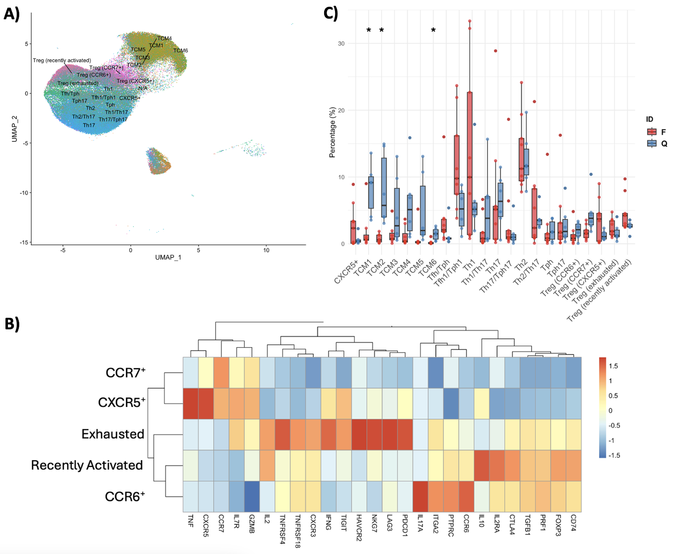
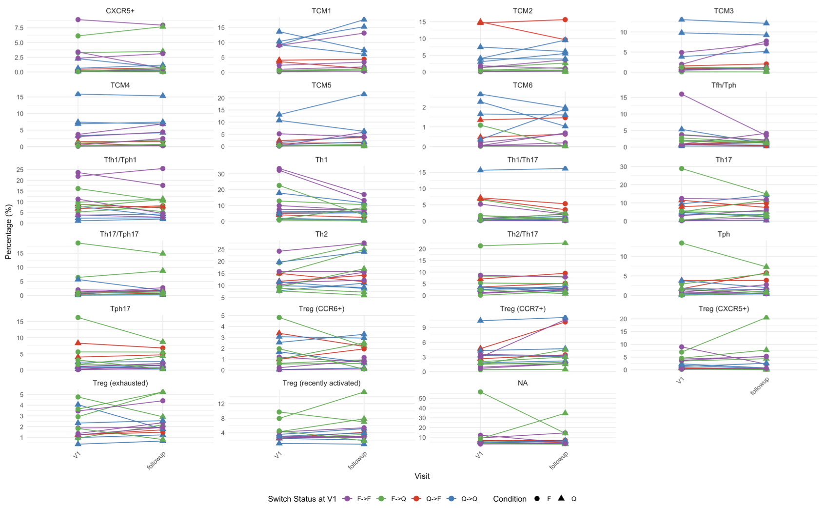
Results: Integration of the gene and surface protein expression data allowed us to identify 23 unique immune populations [Fig.1.A]. Among these, we detected five regulatory T cell (Treg)-enriched clusters expressing canonical Treg-associated genes. Analysis of the relative expression of key molecules among these clusters [Fig.1.B] revealed one subset with high exhaustion marker expression and features consistent with cell reprogramming to a more inflammatory phenotype (increased IFNg and IL-2 expression). Comparing the proportions of this subset between flaring and quiescent patients at baseline [Fig.1.C], we found no differences, suggesting that Treg exhaustion occurs in SLE irrespective of clinical flare status. Although the frequencies were similar, differential gene expression analysis showed significant differences (e.g., IFN-stimulated genes) in flaring and quiescent patients at baseline, including within the exhausted Treg subset. Additionally, samples from quiescent patients were more enriched for central memory T cells (TCMs), specifically TCM1/2/6, compared to flaring patients (p < 0.05) [Fig.1.C]. Conversely, T follicular helper (Tfh), T peripheral helper (Tph), and Th1 cells were more enriched in flaring patients at baseline. Longitudinal analysis demonstrated that TCM proportions remained stable over time [Fig.2], even when patients’ flare status changed between visits.

Conclusion: Treg exhaustion appears consistent regardless of flare status, while differential gene expression highlights the differences between disease states. Quiescent patients show higher enrichment of TCMs, while flaring patients have increased Tfh, Tph, and Th1 cells. These preliminary findings highlight the distinct transcriptional profiles and subset distributions of CD4+ memory T cells in SLE patients, providing insights into the immune mechanisms underlying disease flares and quiescence.
Supporting image 1

Figure 1: A) UMAP depicting 23 annotated clusters. B) Heatmap with z-score normalized mean expression of Treg-associated genes across the Treg-enriched clusters. C) Boxplots showing distribution of proportional percentages of annotated CD4+ memory T cell subsets in SLE patients at baseline (V1). Each dot represents an individual patient’s data point. Patients are grouped by clinical flare status: Flaring (F, red) and Quiescent (Q, blue). To assess for statistical difference, Mann-Whitney U test was used, with p-values adjusted for multiple comparisons using the Benjamini-Hochberg method (*, adjusted p < 0.05).

Supporting image 2

Figure 2: Longitudinal changes in the percentage of CD4+ memory T cell subsets in SLE patients. Proportional percentage of CD4+ memory T cell subsets across two visits (baseline (V1) and follow-up) for each patient. Lines connect individual patient data points, colored by switch status at baseline: Q->F (Quiescent to Flaring), Q->Q (Quiescent to Quiescent), F->Q (Flaring to Quiescent), and F->F (Flaring to Flaring). Points represent individual measurements, with different shapes indicating different conditions, Flaring (F, circle) and Quiescent (Q, triangle). Statistical significance was assessed using the Wilcoxon signed-rank test for paired comparisons, with p-values adjusted for multiple comparisons using the Benjamini-Hochberg method.


Disclosures: C. Nassar: None; R. Quevedo: None; M. Ciudad: None; Z. Faheem: None; K. Manion: None; C. Munoz-Grajales: None; M. Kim: None; D. Gladman: AbbVie, 2, 5, Amgen, 2, 5, AstraZeneca, 2, BMS, 2, Celgene, 2, 5, Eli Lilly, 2, 5, Galapagos, 2, 5, Gilead, 2, 5, Janssen, 2, 5, Novartis, 2, 5, Pfizer, 2, 5, UCB, 2, 5; M. Urowitz: None; Z. Touma: None; T. McGaha: None; J. Wither: AstraZeneca, 1, 2, Pfizer, 5.

To cite this abstract in AMA style:

Nassar C, Quevedo R, Ciudad M, Faheem Z, Manion K, Munoz-Grajales C, Kim M, Gladman D, Urowitz M, Touma Z, McGaha T, Wither J. Characterizing Memory T Cell Subsets Associated with SLE Etiopathogenesis [abstract]. Arthritis Rheumatol. 2024; 76 (suppl 9). https://acrabstracts.org/abstract/characterizing-memory-t-cell-subsets-associated-with-sle-etiopathogenesis/. Accessed .
  • Tweet
  • Email a link to a friend (Opens in new window) Email
  • Print (Opens in new window) Print

« Back to ACR Convergence 2024

ACR Meeting Abstracts - https://acrabstracts.org/abstract/characterizing-memory-t-cell-subsets-associated-with-sle-etiopathogenesis/

Advanced Search

My Favorites

Save and print abstracts during your browser session by clicking the “Favorite” button at the bottom of any abstract (must have cookies enabled in browser). See saved favorites.

Abstract Policies

  • ACR Convergence Abstract Embargo Policies
  • ACR Convergence Abstract Permissions & Reprints
  • PRSYM Abstract Policies

ACR Convergence. Where Rheumatology Meets

ACR Convergence 2026

Join us November 6-11 in Orlando, Florida.
See Registration Information

  • Contact ACR
  • Privacy Policy
  • ACR Policies
  • Cookie Preferences

© Copyright 2026 American College of Rheumatology