Session Information
Session Type: Poster Session A
Session Time: 8:30AM-10:30AM
Background/Purpose: To determine the association between radiographic cam and pincer morphology and loss of hip quantitative joint space width (qJSW) in a community-based cohort.
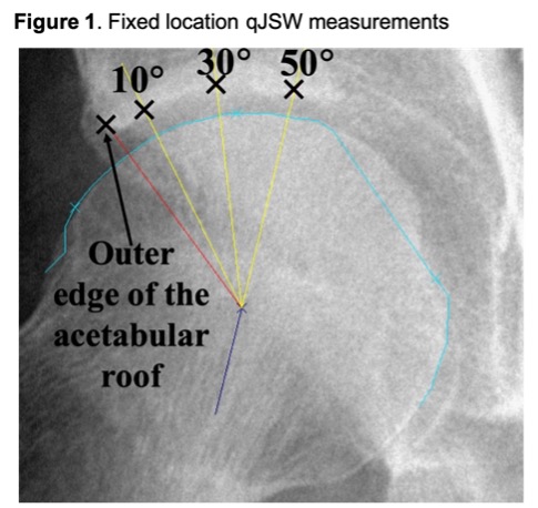
Methods: Data were from Johnston County OA Project (JoCoOA) participants ( >45 years of age) with anteroposterior (AP) pelvis radiographs from baseline and up to 2 follow-up timepoints. A musculoskeletal radiologist assigned all hips a Kellgren-Lawrence grade (KLG). Hip morphology measures were defined on baseline radiographs using OxMorf software (Oxford, UK) by two readers. An independent reader measured qJSW at all timepoints (at 3 locations, Fig 1) using a validated method. All readers were blinded to all other data. Participants self-reported age and race; their height was measured, and body mass index (BMI) calculated at clinic visits. Change in qJSW from baseline to final available follow-up divided by time was modeled by location (10, 30, and 50°) and stratified by sex. Population-averaged linear regression was used to model qJSW change in mm/10 years (mm/10y), accounting for correlation among hips and adjusting for baseline age, race, height, BMI, and qJSW to produce β and 95% confidence intervals [CI]. A sensitivity analysis considered only those hips without baseline rHOA (KLG < 2).
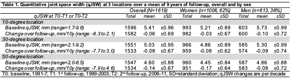
Results: Data from 821 individuals (38% men, 26% Black) with 1619 hips, baseline mean ± SD age 62 ± 9 years, BMI 29 ± 6 kg/m2 and follow-up (at T1 and/or T2; 9 ± 3 years) were included. At baseline, 9% of women and 26% of men had an AP alpha angle >60° consistent with cam morphology. Acetabular under-coverage (i.e., lateral center edge angle [LCEA] £ 25°) was present in about ¼ of the sample and more common in men (31% vs. 22%), while acetabular over-coverage (i.e., LCEA >40° or protrusio) was more common in women (9% vs 5% and 6% vs 0%, respectively). Baseline mean qJSW was around 5mm and decreased by an average of 0.1mm/10y (Table 1).
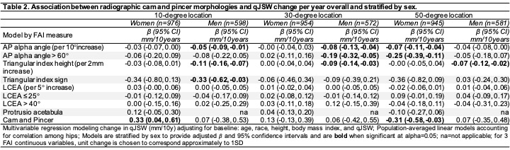
Among women, AP alpha angle and mixed cam/pincer morphology were significantly associated with loss of qJSW at the 50-degree location (up to 0.31mm/10y). In men, indicators of cam morphology (AP alpha angle and triangular index) were associated with loss of qJSW at all 3 locations (up to 0.33mm/10y). Acetabular under- or over-coverage were not significantly associated with change in qJSW in men or women (Table 2).
When considering only 1303 hips without baseline rHOA (data not shown), AP alpha angle was no longer significantly associated with loss of qJSW in women at the 50-degree location, but acetabular overcoverage became significant (-0.22 [-.40, -0.04]mm/10y), and mixed cam/pincer morphology was strongly associated with qJSW loss (-1.20 [-1.67, -0.73]mm/10y). In men, greater triangular index height remained associated with loss of qJSW at most locations (around 0.1mm/10y) but associations with other cam morphologies were not significant.
Conclusion: These data support a role for cam and pincer morphologies in progression of hip joint space loss. Some of these morphologies increased loss of joint space by more than 3 times (e.g., 0.3mm vs 0.1mm/10y) over the baseline average, suggesting that such measures could be used for risk stratification in clinical studies to identify hips at higher risk for progression.
To cite this abstract in AMA style:
Nelson A, Alvarez C, Golightly Y, Stiller J, Renner J, Arden N, Ratzlaff C, Duryea J. Baseline Cam or Pincer Morphology Is Associated with Loss of Quantitative Joint Space Width at the Hip: The Johnston County Osteoarthritis Project [abstract]. Arthritis Rheumatol. 2021; 73 (suppl 9). https://acrabstracts.org/abstract/baseline-cam-or-pincer-morphology-is-associated-with-loss-of-quantitative-joint-space-width-at-the-hip-the-johnston-county-osteoarthritis-project/. Accessed .« Back to ACR Convergence 2021
ACR Meeting Abstracts - https://acrabstracts.org/abstract/baseline-cam-or-pincer-morphology-is-associated-with-loss-of-quantitative-joint-space-width-at-the-hip-the-johnston-county-osteoarthritis-project/