ACR Meeting Abstracts

ACR Meeting Abstracts

  • Meetings
    • ACR Convergence 2024
    • ACR Convergence 2023
    • 2023 ACR/ARP PRSYM
    • ACR Convergence 2022
    • ACR Convergence 2021
    • ACR Convergence 2020
    • 2020 ACR/ARP PRSYM
    • 2019 ACR/ARP Annual Meeting
    • 2018-2009 Meetings
    • Download Abstracts
  • Keyword Index
  • Advanced Search
  • Your Favorites
    • Favorites
    • Login
    • View and print all favorites
    • Clear all your favorites
  • ACR Meetings

Abstract Number: 0848

Automatic Hand Segmentation from Hand X-rays Using Minimized Training Samples and Machine Learning Models

Zilong Yang1, Juan Shan2, Carmine Guida2, Jordan Blackadar1, Tino Cheung1, Jeffrey Driban3, Timothy McAlindon4 and Ming Zhang1, 1Wentworth Institute of Technology, Boston, MA, 2Pace University, New York, NY, 3Tufts Medical Center, Boston, MA, 4Tufts Medical Center, Arlington, MA

Meeting: ACR Convergence 2021

Keywords: Biomarkers, Imaging, Osteoarthritis

  • Tweet
  • Click to email a link to a friend (Opens in new window) Email
  • Click to print (Opens in new window) Print
Session Information

Date: Sunday, November 7, 2021

Title: Research Methodology Poster (0846–0854)

Session Type: Poster Session B

Session Time: 8:30AM-10:30AM

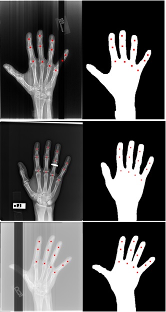
Background/Purpose: In many hand osteoarthritis (OA) studies (e.g., automatic Joint Space Width measuring), accurate hand segmentation is the first step towards making further analysis. However, it would be a difficult task to use only traditional image processing techniques due to inconsistencies like bands, rings, and low contrast X-rays (see Figure 1). To train a machine learning model to segment hand X-rays, one needs to set up a training set in which each hand X-ray should be manually delineated. To minimize the effort of manual delineation, we developed a novel iterative training strategy that requires only a small number of images to be manually segmented.

Methods: 3557 hand X-rays from the Osteoarthritis Initiative (OAI) were utilized in this study, where each X-ray contains one hand. We randomly separated the dataset into a training set with 3024 X-rays and a testing set with 533 X-rays, and the testing set was not exposed to the models during the training procedure. In round 1, we applied a traditional image segmentation algorithm on the training set and selected 132 good-quality results as a training set to train model 1. Round 2, we applied to model 1 to the training set with 3024 images and selected 25 of the worst predicted segmentations from the results. We manually segmented those 25 failed images and used them to train a new U-net model (model 2). In round 3, likewise, we applied to model 2 to the training set again and picked 11 failed segmentations from the results. We manually segmented those 11 failed cases and combined them with the 25 cases from round 2 to train a new model using a total of 36 training samples (model 3). In total, we only manually segmented 36 hand X-rays (25 in round 2 and 11 in round 3). To evaluate segmentation accuracy, two research assistants manually marked the centers of 12 joints (excluding the thumb, Figure 1 red dots) on each of the 533 hands in the testing set. The reason that we used joint centers instead of segmenting the whole hand is to save labor. We evaluated the hand segmentation accuracy at the joint level: if a joint center is within the predicted hand mask, the joint is a match; otherwise, the joint is a miss. The overall accuracy of a model is equal to the number of matches divided by the number of total joints.

Results: Table 1 lists the accuracy for each model: model 1 performed the initial segmentation with the most training samples, but its accuracy was lowest; model 2 used only 25 training samples but improved the accuracy significantly; model 3 achieved the best accuracy (99.95%) with 36 training samples and only 3 joints out of 6396 joints were missed in the testing set.

Conclusion: Acquiring a large medical image dataset is a challenge in many studies and manually labeling is often a time-consuming procedure. In this paper, we proposed an iterative strategy to minimize the training samples that need to be manually labeled. In a dataset of 3557 hand X-rays, we only need to manually segment 36 images and achieved 99.95% accuracy for joint detection on the testing set. This strategy has potential usage in other medical image processing problems to save the manual labeling effort (e.g., knee cartilage segmentation, bone marrow lesion segmentation, etc.).

Figure 1. The first column contains challenge hand X-ray images with white bands, rings, and low contrast. The red dots are manual marks of 12 joint centers. The second column is the predicted hand masks by the machine learning model U-net (model 3).

Table 1: Hand segmentation accuracy


Disclosures: Z. Yang, None; J. Shan, None; C. Guida, None; J. Blackadar, None; T. Cheung, None; J. Driban, None; T. McAlindon, Kolon Tissue Gene, 1, Organogenesis, 1, Pfizer Inc., 1, Regeneron, 1, Remedium Bio, 1, Samumed, 1, Seikagaku, 1; M. Zhang, None.

To cite this abstract in AMA style:

Yang Z, Shan J, Guida C, Blackadar J, Cheung T, Driban J, McAlindon T, Zhang M. Automatic Hand Segmentation from Hand X-rays Using Minimized Training Samples and Machine Learning Models [abstract]. Arthritis Rheumatol. 2021; 73 (suppl 9). https://acrabstracts.org/abstract/automatic-hand-segmentation-from-hand-x-rays-using-minimized-training-samples-and-machine-learning-models/. Accessed .
  • Tweet
  • Click to email a link to a friend (Opens in new window) Email
  • Click to print (Opens in new window) Print

« Back to ACR Convergence 2021

ACR Meeting Abstracts - https://acrabstracts.org/abstract/automatic-hand-segmentation-from-hand-x-rays-using-minimized-training-samples-and-machine-learning-models/

Advanced Search

Your Favorites

You can save and print a list of your favorite abstracts during your browser session by clicking the “Favorite” button at the bottom of any abstract. View your favorites »

All abstracts accepted to ACR Convergence are under media embargo once the ACR has notified presenters of their abstract’s acceptance. They may be presented at other meetings or published as manuscripts after this time but should not be discussed in non-scholarly venues or outlets. The following embargo policies are strictly enforced by the ACR.

Accepted abstracts are made available to the public online in advance of the meeting and are published in a special online supplement of our scientific journal, Arthritis & Rheumatology. Information contained in those abstracts may not be released until the abstracts appear online. In an exception to the media embargo, academic institutions, private organizations, and companies with products whose value may be influenced by information contained in an abstract may issue a press release to coincide with the availability of an ACR abstract on the ACR website. However, the ACR continues to require that information that goes beyond that contained in the abstract (e.g., discussion of the abstract done as part of editorial news coverage) is under media embargo until 10:00 AM ET on November 14, 2024. Journalists with access to embargoed information cannot release articles or editorial news coverage before this time. Editorial news coverage is considered original articles/videos developed by employed journalists to report facts, commentary, and subject matter expert quotes in a narrative form using a variety of sources (e.g., research, announcements, press releases, events, etc.).

Violation of this policy may result in the abstract being withdrawn from the meeting and other measures deemed appropriate. Authors are responsible for notifying colleagues, institutions, communications firms, and all other stakeholders related to the development or promotion of the abstract about this policy. If you have questions about the ACR abstract embargo policy, please contact ACR abstracts staff at [email protected].

Wiley

  • Online Journal
  • Privacy Policy
  • Permissions Policies
  • Cookie Preferences

© Copyright 2025 American College of Rheumatology