ACR Meeting Abstracts

ACR Meeting Abstracts

  • Meetings
    • ACR Convergence 2024
    • ACR Convergence 2023
    • 2023 ACR/ARP PRSYM
    • ACR Convergence 2022
    • ACR Convergence 2021
    • ACR Convergence 2020
    • 2020 ACR/ARP PRSYM
    • 2019 ACR/ARP Annual Meeting
    • 2018-2009 Meetings
    • Download Abstracts
  • Keyword Index
  • Advanced Search
  • Your Favorites
    • Favorites
    • Login
    • View and print all favorites
    • Clear all your favorites
  • ACR Meetings

Abstract Number: 1231

[68Ga]- CPCR4 Trifluoroacetate PET/CT Imaging of Chemokine Receptor CXCR4- Expression in Systemic Sclerosis Related Interstitial Lung Disease

Chirag Rajkumar Kopp1, Shefali Sharma2, Venkata Subramanian Krishnaraju3, Chandra Bhushan Prasad3, Jagdeep Singh3, Shashi Anand3, Ranjana Minz3, Ashwani Sood3, Anindita Sinha3, Rajender Kumar Basher3, Sahajal Dhooria3, Varun Dhir4 and sanjay jain3, 1Post Graduate Institute of Medical Education and Research, Chandigarh, India, 2PGIMER< Chandigarh, Chandigarh, India, 3Postgraduate Institute of Medical Education and Research, Chandigarh, India, 4PGIMER, CHD, INDIA, Chandigarh, India

Meeting: ACR Convergence 2022

Keywords: Computed tomography (CT), Imaging, immunology, interstitial lung disease, Systemic sclerosis

  • Tweet
  • Click to email a link to a friend (Opens in new window) Email
  • Click to print (Opens in new window) Print
Session Information

Date: Sunday, November 13, 2022

Title: Imaging of Rheumatic Diseases Poster

Session Type: Poster Session C

Session Time: 1:00PM-3:00PM

Background/Purpose: Interstitial lung disease (ILD) in Systemic sclerosis (SSc) is a major contributor of morbidity and mortality. CXCR4/CXCL12 axis has been implicated in the pathogenesis of pulmonary fibrosis. In this pilot study, we characterize the CXCR4 expression in lung parenchyma using 68Ga-Lympholeucin (CPCR4 trifluoroacetate) and peripheral white blood cells using PET/CT and flow cytometry.

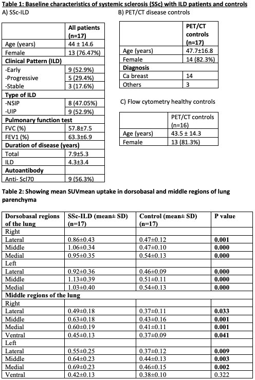
Methods: In this cross-sectional study, we included SSc patients having active (n=14) and stable(n=3) ILD based on the duration, clinical and radiological progression, and FVC decline. 68Ga-Lympholeucin uptake in lung parenchyma was quantified in these 17 SSc-ILD patients and compared to the diseased controls without lung parenchymal involvement. Lung parenchyma was divided into 22 regions of interest (ROI) on PET/CT, drawn in the dorsobasal, middle and apical regions of the lung and individual areas were quantified for CXCR4 expression using SUVmean and compared to the controls. Flow cytometry was performed in 16 patients to quantify CXCR4 expression on CD4+ T cell, CD8+ T cell, CD14+ monocytes and CD19+ B cells, and compared with healthy controls.

Results: SSc-ILD patients underwent 68Ga-Lympholeucin PET/CT imaging showed significant SUVmean uptake in the lung parenchyma compared to controls. Most significant areas of uptake were noted in the dorsobasal regions of lung. Also, significantly higher CXCR4 uptake in the areas of ground glass (P< 0.001), reticulation (P< 0.001) and reticulation with architectural distortion (P< 0.001) opacities as compared to normal regions. Higher proportion of CD4 (83.83±7.99 vs 60.28±12.20; P< 0.001) and CD8 (67.67±14.4 vs 43.67±12.59; P=0.003) cells in SSc-ILD patients showed CXCR4 expression as compared to controls.

Conclusion: 68Ga-Lympholeucin PET/CT is a good imaging modality to quantify CXCR4 expression in lung parenchyma of SSc-ILD patients. Increased CXCR4 expression in the lung parenchyma using PET/CT and peripheral T cells could be used as a biomarker of disease activity and assessment of response to therapy.

Supporting image 1

Supporting image 2

In patient with stable disease, transaxial CT images (A) showing reticular opacities with subpleural sparing and areas of architectural distortion with increased tracer uptake on the fused PET/CT images (B). Reformatted sagittal CT (C) and fused PET/CT (D) images showing the changes confined predominantly to the basal zones with relative sparing of the upper lobes. In patient with active disease, (E) showing reticular opacities and areas of patchy consolidation and ground glassing with increased tracer uptake on the fused PET/CT images (F). Reformatted sagittal CT (G) and fused PET/CT (H) images showing similar changes predominantly in the posterior lung fields with focal areas of architectural distortion
In control patient, transaxial (I) and reformatted sagittal CT images (J) showing no abnormality on the bilateral lung fields with no abnormal tracer uptake in the lung fields on the fused PET/CT images (K,L).

Supporting image 3

Representative flow diagrams showing gating strategy for CXCR4 expression: a) Gating of lymphocytes on FSC and SSC dot plot and monocytes were gated on cluster present between lymphocytes and granulocytes , b) showing CD3+ T cells as P2, CD19+ B cells as P3, c) showing CD4+ T cells as P4 and CD4- CD3+ (CD8 T cells) as P5 d) Showing CD14+ monocytes as P9, (e,f,g,h) showing CXCR4 expression on CD4+ T cells as p6, CD8+ T cells as p7, CD19+ B cells as p8 and CD14+ monocytes as p 10, respectively.


Disclosures: C. Kopp, None; S. Sharma, None; V. Krishnaraju, None; C. Prasad, None; J. Singh, None; S. Anand, None; R. Minz, None; A. Sood, None; A. Sinha, None; R. Basher, None; S. Dhooria, None; V. Dhir, None; s. jain, None.

To cite this abstract in AMA style:

Kopp C, Sharma S, Krishnaraju V, Prasad C, Singh J, Anand S, Minz R, Sood A, Sinha A, Basher R, Dhooria S, Dhir V, jain s. [68Ga]- CPCR4 Trifluoroacetate PET/CT Imaging of Chemokine Receptor CXCR4- Expression in Systemic Sclerosis Related Interstitial Lung Disease [abstract]. Arthritis Rheumatol. 2022; 74 (suppl 9). https://acrabstracts.org/abstract/68ga-cpcr4-trifluoroacetate-pet-ct-imaging-of-chemokine-receptor-cxcr4-expression-in-systemic-sclerosis-related-interstitial-lung-disease/. Accessed .
  • Tweet
  • Click to email a link to a friend (Opens in new window) Email
  • Click to print (Opens in new window) Print

« Back to ACR Convergence 2022

ACR Meeting Abstracts - https://acrabstracts.org/abstract/68ga-cpcr4-trifluoroacetate-pet-ct-imaging-of-chemokine-receptor-cxcr4-expression-in-systemic-sclerosis-related-interstitial-lung-disease/

Advanced Search

Your Favorites

You can save and print a list of your favorite abstracts during your browser session by clicking the “Favorite” button at the bottom of any abstract. View your favorites »

All abstracts accepted to ACR Convergence are under media embargo once the ACR has notified presenters of their abstract’s acceptance. They may be presented at other meetings or published as manuscripts after this time but should not be discussed in non-scholarly venues or outlets. The following embargo policies are strictly enforced by the ACR.

Accepted abstracts are made available to the public online in advance of the meeting and are published in a special online supplement of our scientific journal, Arthritis & Rheumatology. Information contained in those abstracts may not be released until the abstracts appear online. In an exception to the media embargo, academic institutions, private organizations, and companies with products whose value may be influenced by information contained in an abstract may issue a press release to coincide with the availability of an ACR abstract on the ACR website. However, the ACR continues to require that information that goes beyond that contained in the abstract (e.g., discussion of the abstract done as part of editorial news coverage) is under media embargo until 10:00 AM ET on November 14, 2024. Journalists with access to embargoed information cannot release articles or editorial news coverage before this time. Editorial news coverage is considered original articles/videos developed by employed journalists to report facts, commentary, and subject matter expert quotes in a narrative form using a variety of sources (e.g., research, announcements, press releases, events, etc.).

Violation of this policy may result in the abstract being withdrawn from the meeting and other measures deemed appropriate. Authors are responsible for notifying colleagues, institutions, communications firms, and all other stakeholders related to the development or promotion of the abstract about this policy. If you have questions about the ACR abstract embargo policy, please contact ACR abstracts staff at [email protected].

Wiley

  • Online Journal
  • Privacy Policy
  • Permissions Policies
  • Cookie Preferences

© Copyright 2025 American College of Rheumatology