Session Information
Session Type: Abstract Submissions (ACR)
Background/Purpose: We and others have demonstrated low grade inflammation exists in OA joint tissues, where it may contribute to disease pathogenesis. In the current studies we assessed whether inflammatory events occurring within joint tissues were reported in the peripheral blood leukocytes (PBLs) of patients with symptomatic knee OA (SKOA).
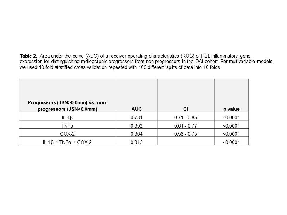
Methods: PBL inflammatory gene expression (IL-1, TNFα, COX-2) was assessed in two independent cohorts of patients with SKOA, and a cohort of healthy control subjects: 1) 111 patients with tibiofemoral medial OA and 21 healthy volunteers from the NYUHJD Cohort, and 2) 200 patients from the OAI progression cohort who had “high quality radiographs”, at both baseline and 24 months, and had KL2 or 3 in the signal knee at baseline. Radiographic progression was defined as narrowing of medial joint space width (JSW) in the signal knee between baseline and 24-months in each cohort. Fast progressors were defined as subjects who had JSN >0.5mm over 24 months. For measuring predictive performance, we used the area under the curve (AUC) of a receiver operating characteristics (ROC). OAI SKOA subjects were dichotomized as radiographic non-progressors (JSN <0.0 mm) and progressors (JSN>0.0mm) for association studies.
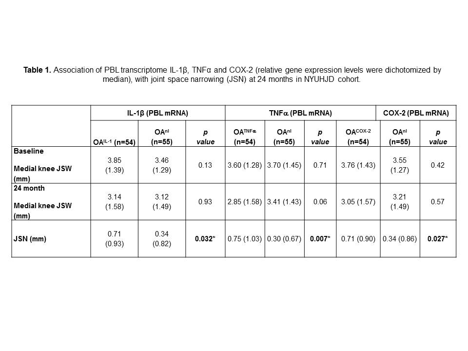
Results: Elevated PBL expression of IL-1, TNFα or COX-2 at baseline identified SKOA patients who were “fast progressors” (mean JSN 0= 0.71, 0.75 and 0.71 mm / 24 months, respectively) compared to patients with levels below the median (Table 1). In a multivariable model, anthropometric traits alone (BMI, gender, age) did not predict progression, whereas addition of PBL gene expressions improved prediction of fast progressors (JSN>0.5mm). We next examined inflammatory gene expression in PBLs of radiographic progressors in the OAI cohort. Similar to the NYUHJD cohort, elevated expression of IL-1β, TNFα and COX-2 mRNA at baseline distinguished radiographic progressors from non-progressors (Table 2).
Conclusion: We identified, and confirmed in two cohorts, increased inflammatory gene expression (IL-1, TNFα or COX-2) by PBLs that predict radiographic progression in patients with SKOA. The data indicate that inflammatory events within joint tissues of patients with SKOA are reported in the peripheral blood. These PBL transcriptome signals of local joint inflammation merit further study as potential biomarkers for OA disease progression.
Disclosure:
M. Attur,
Patent,
9;
A. Statnikov,
None;
S. Krasnokutsky Samuels,
None;
V. B. Kraus,
NIAMS-NIH,
2;
J. Jordan,
None;
B. D. Mitchell,
None;
M. Yau,
None;
J. Patel,
None;
C. F. Aliferis,
None;
M. C. Hochberg,
NIH,
2;
J. Samuels,
None;
S. B. Abramson,
NIAMS-NIH,
2,
Patent,
9.
« Back to 2014 ACR/ARHP Annual Meeting
ACR Meeting Abstracts - https://acrabstracts.org/abstract/elevated-peripheral-blood-leukocyte-inflammatory-gene-expression-in-radiographic-progressors-with-symptomatic-knee-osteoarthritis-nyu-and-oai-cohorts/


