Session Information
Session Type: Abstract Submissions (ACR)
Background/Purpose: Osteophytes are cartilage capped, bony outgrowths that limit joint movement and originate from the periosteum or from ligaments during osteoarthritis (OA). There is increasing belief that the synovium contributes to OA joint pathology since a large subset of OA patients shows synovial activation. As shown recently in our lab, alarmins S100A8 and S100A9 (major products of activated synovial macrophages) are involved in cartilage degradation and synovial activation during human and murine OA. In the current study, we explored the involvement of S100A8/A9 in osteophyte formation in experimental OA.
Methods: Experimental OA was elicited in C57Bl/6 (WT) mice and S100A9-/- mice (in which peripheral myeloid cells lack functional S100A8), either by intra-articular collagenase injection (CIOA), or by transsection of the medial anterior meniscotibial ligament (DMM). Osteophyte size was assessed by a blinded observer using image analysis software. Chondrogenesis was induced by bringing human fetal mesenchymal stem cells (hMSCs) in pellet or murine C3H10T1/2 in micromass culture and stimulating for 5 (hMSCs) or 21 days (C3H10T1/2) with BMP-2 and TGFβ1, with 1 or 5 µg/ml human or mouse recombinant S100A8. Proteoglycan content was quantified on SafO stained sections, expression of mRNA with RT-qPCR.
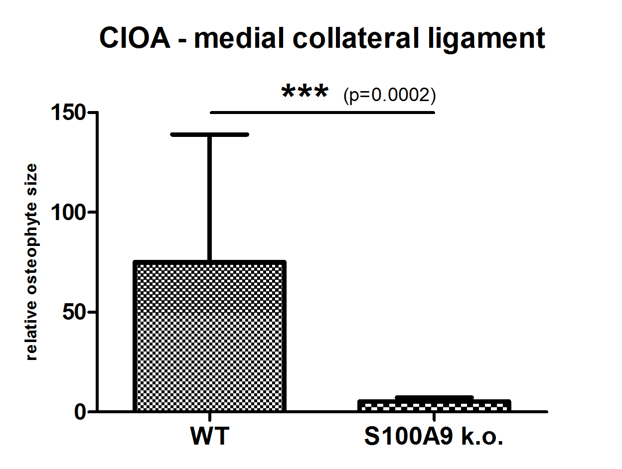
Results: Synovial activation, which is high in CIOA, was significantly reduced in S100A9-/- mice. Osteophyte size at day 42 of CIOA was dramatically reduced in the S100A9-/- compared to WT in the medial collateral ligament (92,5% reduction, Figure 1), but also significantly at the medial side of both tibia and femur (68,2% and 64,6% reduction) (n=10). One explanation for the reduced osteophyte size in S100A9-/- mice may be a direct effect of S100-proteins on chondrogenesis. To investigate this, we first stimulated murine C3H10T1/2 MSCs in micromass culture with 5 µg/ml S100A8 (in the presence of BMP-2 and TGFβ1) and found a marked increase in MMP3 and aggrecan mRNA as well as a strongly altered morphology, indicating increased remodeling. In line with that, stimulation of human MSCs in pellet culture with 1 or 5 µg/ml S100A8 (again together with BMP-2 and TGFβ1) strongly increased proteoglycan deposition as measured by redness in SafO staining (27% and 71% increase respectively). Finally, we determined osteophyte size in the DMM model, in which synovial involvement is very low. At day 56, we observed no significant differences in osteophyte size between the S100A9-/- and WT at the medial femur and tibia (105% and 136% of WT, n=8).
Conclusion: S100A8/S100A9 play a crucial role in osteophyte formation in an OA model that shows high synovial involvement, probably by stimulating chondrogenesis. Considering also the deleterious effect of S100A8/A9 on joint destruction in OA, targeting these alarmins during OA may be very promising.
Disclosure:
R. Schelbergen,
None;
A. B. Blom,
None;
W. de Munter,
None;
A. W. Sloetjes,
None;
T. Vogl,
None;
J. Roth,
None;
W. B. van den Berg,
None;
P. L. E. M. van Lent,
None.
« Back to 2013 ACR/ARHP Annual Meeting
ACR Meeting Abstracts - https://acrabstracts.org/abstract/alarmins-s100a8a9-regulate-osteophyte-formation-in-experimental-osteoarthritis-with-high-synovial-activation/

