Session Information
Date: Monday, October 27, 2025
Title: (0934–0954) Systemic Lupus Erythematosus – Animal Models Poster
Session Type: Poster Session B
Session Time: 10:30AM-12:30PM
Background/Purpose: Lupus is a heterogeneous inflammatory autoimmune disease affecting a variety of organs, including skin manifestations such as photosensitivity and cutaneous lesions. The skin of human lupus patients is primed for disease through a variety of cellular mechanisms, including an increase in Type I interferons (IFNs) such as interferon kappa (IFNk) that activate immune cells and prime for photosensitivity. Here, we study the effects of epidermal transgenic (Tg) Ifnk overexpression to determine the role of skin-derived type I IFNs in driving lupus phenotypes. This experimental work utilized mice of C57/Bl6 background.
Methods: TgIFNk mice were generated utilizing the K14 promoter to direct Ifnk expression to the basal epidermis as previously reported. Mice were monitored for spontaneous lesions in a 12-month survival cohort. Where indicated, 3-month-old mice were exposed to 300mJ/cm2 UVB 12 hours before euthanasia. Phenotypes of TgIFNk and wild type C57/Bl6 mice were studied via complete blood count (CBC), ELISA, flow cytometry, immunohistochemistry (IHC), immunofluorescence (IF), and quantitative real-time polymerase chain reaction (qrt-PCR) performed on blood, spleen, skin, kidney, and urine harvested at time of euthanasia. All data analysis was performed using GraphPad Prism 10, including two-way ANOVA, one-way ANOVA, or unpaired t-Test where appropriate.
Results: In the C57/Bl6 background, only 4 percent of TgIFNk mice developed spontaneous skin lesions, and no mice developed proteinuria. Lesional mice demonstrated elevated circulating leukocytes compared to age-matched sex-matched non-lesional TgIFNk and WT controls. This increase was driven primarily by an expansion of neutrophils. In the skin, however, immunohistochemistry revealed that skin lesions were enriched primarily in monocytes and monocyte-derived dendritic cells. Non-lesional skin showed no change in CD11b+ cells between genotypes, suggesting that increased circulating neutrophils drive enhanced recruitment of myeloid cells upon cutaneous lesion formation. WT and TgIFNk mice undergoing UVB irradiation exhibited increased monocytes in the skin, but no difference was noted between strains.
Conclusion: TgIFNk mice on the C57/Bl6 background exhibit increases in circulating inflammatory innate immune cells and aggressive myeloid-driven skin disease in a minor subset of mice. Future work seeks to further characterize the function of these innate immune cells in both spontaneous lesions and UVB-mediated disease activation.
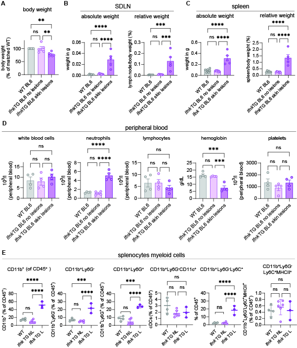 Organ weights (1A-C), Complete blood count (1D), and flow cytometry (1E) of mice generating spontaneous lesions (IfnkTG BL6 skin lesions), and age-matched sex-matched non-lesional TG or wild type (WT) controls illustrating lymphadenopathy, splenomegaly, anemia, increased circulating neutrophils, and increased circulating myeloid cells.
.jpg) Immunohistochemistry (IHC) of CD11b and Ly6G for spontaneous lesions (IfnkTG BL6 skin lesions), and age-matched sex-matched non-lesional TG or wild type (WT) controls.
Immunohistochemistry (IHC) of CD11b and Ly6G for spontaneous lesions (IfnkTG BL6 skin lesions), and age-matched sex-matched non-lesional TG or wild type (WT) controls.
.jpg) UVB experiment graphical abstract and flow cytometry results showing a reduction in monocytes in the spleen compared to an increase in monocytes in the skin.
UVB experiment graphical abstract and flow cytometry results showing a reduction in monocytes in the spleen compared to an increase in monocytes in the skin.
To cite this abstract in AMA style:
O'Brien P, Klein B, Colesa D, Kahlenberg J. Epidermal IFNκ Increases Circulating and Cutaneous Monocytes in a C57/Bl6 Overexpression Mouse Model [abstract]. Arthritis Rheumatol. 2025; 77 (suppl 9). https://acrabstracts.org/abstract/epidermal-ifn%ce%ba-increases-circulating-and-cutaneous-monocytes-in-a-c57-bl6-overexpression-mouse-model/. Accessed .« Back to ACR Convergence 2025
ACR Meeting Abstracts - https://acrabstracts.org/abstract/epidermal-ifn%ce%ba-increases-circulating-and-cutaneous-monocytes-in-a-c57-bl6-overexpression-mouse-model/