ACR Meeting Abstracts

ACR Meeting Abstracts

  • Meetings
    • ACR Convergence 2024
    • ACR Convergence 2023
    • 2023 ACR/ARP PRSYM
    • ACR Convergence 2022
    • ACR Convergence 2021
    • ACR Convergence 2020
    • 2020 ACR/ARP PRSYM
    • 2019 ACR/ARP Annual Meeting
    • 2018-2009 Meetings
    • Download Abstracts
  • Keyword Index
  • Advanced Search
  • Your Favorites
    • Favorites
    • Login
    • View and print all favorites
    • Clear all your favorites
  • ACR Meetings

Abstract Number: 1664

Identification of Spatial Determinants of Treatment Response in Rheumatoid Arthritis Synovia

ilya Korsunsky1, Roopa Madhu2, Kevin Wei3, Kartik Bhamidipati1, Miles Tran1, Sonia Presti1, Anna Helena Jonsson4, Ellen Gravallese5, Michael Brenner6, Soumya Raychaudhuri1, Jennifer Seifert7, Costantino Pitzalis8, Fan Zhang9, Larry Moreland10, V. Michael Holers10, Michael Clayman11, Myles Lewis12, Ce Gao1, Michael J Zuscik13 and Accelerating Medicines Partnership Autoimmune and Immune-Mediated Disease14, 1Brigham and Women's Hospital, Boston, MA, 2Brigham and Women's Hospital, Brookline, MA, 3Brigham and Women's Hospital at Harvard Medical School, Boston, MA, 4University of Colorado School of Medicine, Aurora, CO, 5Brigham and Women's Hospital, Harvard Medical School, Chestnut Hill, MA, 6Brigham and Women's Hospital, Harvard Medical School, Newton, MA, 7University of Colorado and Oklahoma Medical Research Foundation, Aurora, CO, 8QMUL, Bromley Kent, United Kingdom, 9University of Colorado, Aurora, CO, 10University of Colorado, Denver, CO, 11University of Colorado School of Medicine, Burlington, MA, 12Queen Mary University of London, London, United Kingdom, 13Department of Orthopedic Surgery, University of Colorado School of Medicine, Aurora, CO, 14Multiple Institutions, Oklahoma City

Meeting: ACR Convergence 2024

Keywords: autoimmune diseases, Inflammation, rheumatoid arthritis

  • Tweet
  • Click to email a link to a friend (Opens in new window) Email
  • Click to print (Opens in new window) Print
Session Information

Date: Sunday, November 17, 2024

Title: Abstracts: RA – Etiology and Pathogenesis I

Session Type: Abstract Session

Session Time: 1:00PM-2:30PM

Background/Purpose: Rheumatoid arthritis (RA) is an autoimmune disease characterized by immune infiltration of the synovial tissue, resulting in joint damage. In RA, synovia exhibit distinct cellular compositions (CTAP) (1) and variable responses to biologics. We hypothesize that RA synovia comprises distinct cellular niches, and these niches underlie synovial tissue heterogeneity and drive differential treatment response. To this end, we applied spatial transcriptomics to build a spatial atlas of RA synovia

Methods: Spatial transcriptomic profiling

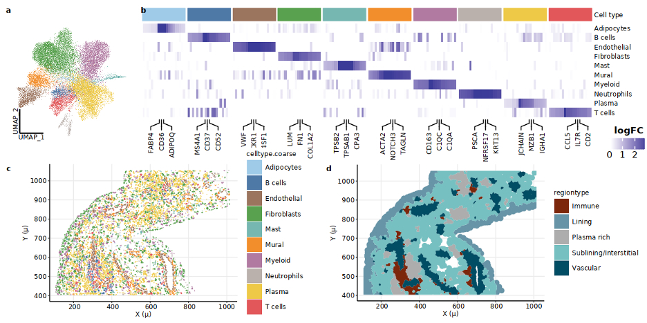
We profiled RA synovial tissue samples (n=10) using CosMx to 1) define synovial cell states and 2) identify cellular regions. Next, we validated novel cellular regions identified in this dataset using Xenium on a separate RA cohort (n=7)

Cell segmentation and niche annotation

We developed a segmentation pipeline to group transcripts into cells and classified each cell into cell types, followed by a label transfer method to define fine-grained cell states. We used a graph-diffusion-based method to identify transcriptionally similar neighborhoods.

Results: Cellular niches in RA synovia.  By applying spatial transcriptomic analysis followed by cellular niche analysis, we identified five spatially distinct regions:(a) lining enriched in lining fibroblasts and MERTK+ HBEGF+ fibroblasts, (b) sublining (sublining fibroblasts and myeloid cells), (c) vascular regions (endothelial and mural cells), (d) plasma cell-rich regions (plasma cells and fibroblasts), and (e) lymphoid aggregates (Fig. 1c-e).


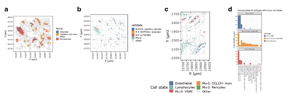
Identification of CCL19+ mural cells.  Analysis of the perivascular niches revealed 3 distinct mural cell types: vascular smooth muscle cells (VSMC), pericytes, and CCL19-expressing CCL19+ mural cells (Fig. 2a). We mapped these cells in situ and identified CCL19+ mural cells are expanded near immune-infiltrated sections of synovial endothelia (Fig 2c). Colocalization analysis revealed that CCL19+ mural cells are spatially associated with T peripheral helper (Tph) cells, age-associated B cells (ABCs), and LAMP3+ DCs (Fig. 2d).


Cellular niches associated with treatment response. We defined two types of sublining niches: leukocyte-rich and leukocyte-poor. Leuko-rich niches are enriched in inflammatory DC3s, MERTK+ SELENOP+ macrophages, DDK3+ fibroblasts, NOTCH3+ fibroblasts, and CD74-hi fibroblasts, while leuko-poor subregions are enriched in CXCL12+ SFRP1+ and POSTN+ fibroblasts, and inflammatory IL1B+ FCN1+ macrophages (Fig. 3). In an independent clinical dataset of bulk RNA-seq from the R4RA trial, we found the gene signatures for leukocyte-rich enriched populations consistently overexpressed in synovial samples from responders (vs non-responders), both before and after treatment and for both Rituximab or Tocilizumab (Fig. 4 and5). 

Conclusion: We established the first spatial atlas of RA synovia at a single-cell resolution. Identifying cellular niches enriched in responders and non-responders to biologics will improve our understanding of cellular and molecular pathways driving treatment outcomes in RA.

Supporting image 1

Spatial architecture of inflamed synovium (a) Cells projected in UMAP space, colored by their major cell type. (b) Heatmap of gene log fold-changes for each of the major cell types. The top 3 marker genes for each cluster are annotated on the x-axis. (c) Cells in tissue space in active RA synovium. () Regions in space: Tissue in a FOV colored by its region type. FOV: Field of view

Supporting image 2

CCL19+ mural cells are enriched near immune infiltrated veins(a) Subtypes of vascular regions highlighted in a FOV (b) Cells belonging to the vascular regions in the FOV shown in (a) colored by their endothelial cell state identity. (c) Cells belonging to the vascular regions in the FOV are shown in (a) colored by their mural cell state identity, and T and B cells are labeled as lymphocytes. (d) Colocalization z-scores of each mural cell state with colocalizing cells.

Supporting image 3

Cell types enriched in Leuko-rich niche are significantly enriched in responders (A) (a) Fraction of myeloid cell states in leuko-poor vs lueko-rich sublining. (b) Fraction of fibroblast cell states in leuko-poor vs lueko-rich sublining. Only cell states that are significantly different between the two types of sublining are highlighted.
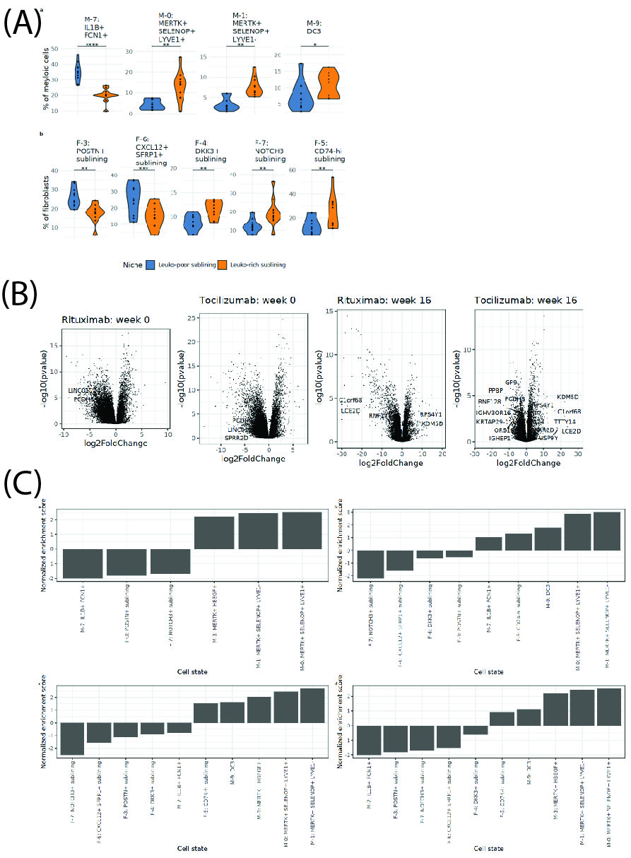
(B) Gene expression in responsders vs non-responders. Volcano plot of differential expression genes (responders vs non-responders) for Rituximab and Tocilizumab at two time points for two different drugs.
(C) Normalized enrichment scores (NES) of the transcriptional profile of cell states in non-responders. (a) NES for cell state transcriptional profiles in patients treated with Rituximab at week 0. (b) NES for cell state transcriptional profiles in patients treated with Tocilizumab at week 0. (c) NES for cell state transcriptional profiles in patients treated with Rituximab at week 16. (d) NES for cell state transcriptional profiles in patients treated with Tocilizumab at week 16.


Disclosures: i. Korsunsky: Mestag Therapeutics LTD, 2; R. Madhu: None; K. Wei: 10X Genomics, 5, Gilead sciences, 2, Merck/MSD, 5, Mestag, 2, santa ana bio, 2; K. Bhamidipati: None; M. Tran: None; S. Presti: None; A. Jonsson: Pfizer, 6; E. Gravallese: Beam Therapeutics, 11, CRISPR Therapeutics, 11, Editas Medicine, 11, Intellia Therapeutics, 11, New England Journal of Medicine, 3, NIH, 5, Rheumatology Research Foundation, 5; M. Brenner: GlaxoSmithKlein(GSK), 2, Mestag Therapeutics, 2, 11, Moderna, 2; S. Raychaudhuri: Janssen, 1, Mestag, 8, Nimbus, 2, Pfizer, 1, Sonoma, 8, Third Rock Ventures, 2; J. Seifert: None; C. Pitzalis: AbbVie/Abbott, 2, 5, 6, AnaptysBio, 2, 5, Exagen, 2, Janssen, 2, 5, 6, Kinikska, 2, Novartis, 2, 5, Pfizer, 5, Sanofi, 2, 5, 6; F. Zhang: None; L. Moreland: None; V. Holers: None; M. Clayman: None; M. Lewis: None; C. Gao: None; M. Zuscik: None; A. Autoimmune and Immune-Mediated Disease: None.

To cite this abstract in AMA style:

Korsunsky i, Madhu R, Wei K, Bhamidipati K, Tran M, Presti S, Jonsson A, Gravallese E, Brenner M, Raychaudhuri S, Seifert J, Pitzalis C, Zhang F, Moreland L, Holers V, Clayman M, Lewis M, Gao C, Zuscik M, Autoimmune and Immune-Mediated Disease A. Identification of Spatial Determinants of Treatment Response in Rheumatoid Arthritis Synovia [abstract]. Arthritis Rheumatol. 2024; 76 (suppl 9). https://acrabstracts.org/abstract/identification-of-spatial-determinants-of-treatment-response-in-rheumatoid-arthritis-synovia/. Accessed .
  • Tweet
  • Click to email a link to a friend (Opens in new window) Email
  • Click to print (Opens in new window) Print

« Back to ACR Convergence 2024

ACR Meeting Abstracts - https://acrabstracts.org/abstract/identification-of-spatial-determinants-of-treatment-response-in-rheumatoid-arthritis-synovia/

Advanced Search

Your Favorites

You can save and print a list of your favorite abstracts during your browser session by clicking the “Favorite” button at the bottom of any abstract. View your favorites »

All abstracts accepted to ACR Convergence are under media embargo once the ACR has notified presenters of their abstract’s acceptance. They may be presented at other meetings or published as manuscripts after this time but should not be discussed in non-scholarly venues or outlets. The following embargo policies are strictly enforced by the ACR.

Accepted abstracts are made available to the public online in advance of the meeting and are published in a special online supplement of our scientific journal, Arthritis & Rheumatology. Information contained in those abstracts may not be released until the abstracts appear online. In an exception to the media embargo, academic institutions, private organizations, and companies with products whose value may be influenced by information contained in an abstract may issue a press release to coincide with the availability of an ACR abstract on the ACR website. However, the ACR continues to require that information that goes beyond that contained in the abstract (e.g., discussion of the abstract done as part of editorial news coverage) is under media embargo until 10:00 AM ET on November 14, 2024. Journalists with access to embargoed information cannot release articles or editorial news coverage before this time. Editorial news coverage is considered original articles/videos developed by employed journalists to report facts, commentary, and subject matter expert quotes in a narrative form using a variety of sources (e.g., research, announcements, press releases, events, etc.).

Violation of this policy may result in the abstract being withdrawn from the meeting and other measures deemed appropriate. Authors are responsible for notifying colleagues, institutions, communications firms, and all other stakeholders related to the development or promotion of the abstract about this policy. If you have questions about the ACR abstract embargo policy, please contact ACR abstracts staff at [email protected].

Wiley

  • Online Journal
  • Privacy Policy
  • Permissions Policies
  • Cookie Preferences

© Copyright 2025 American College of Rheumatology