Session Information
Session Type: Poster Session C
Session Time: 9:00AM-11:00AM
Background/Purpose: To identify features of cartilage thickness change and baseline clinical indicators associated with the transition from normal to mildly reduced joint space as an early indicator of structural knee osteoarthritis (KOA).
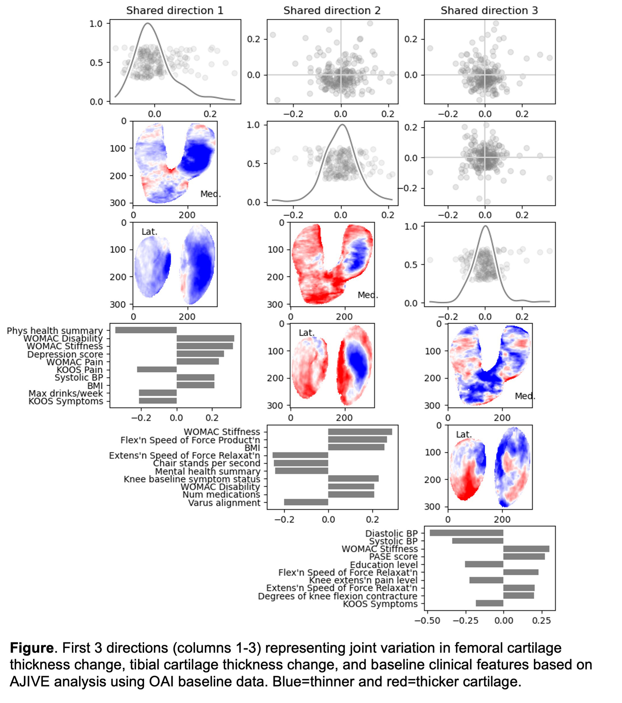
Methods: We identified knees with radiographic medial joint space narrowing (JSN) grade 0 that transitioned to JSN grade ≥1 between any two timepoints during 96 months of follow-up in the Osteoarthritis Initiative (OAI, n=349) and selected cartilage maps (using a 3D U-Net as described in 10.1016/j.ocarto.2023.100334) from the last visit with JSN=0 and the first visit with JSN≥1. Difference maps reflecting the change in cartilage thickness at this transition for the femur and for the tibia were generated and used as data blocks. Angle-based Joint and Individual Variation Explained (AJIVE) was applied to identify the modes of variation expressed in 3 data blocks: (i) femoral cartilage change maps (ii) tibial cartilage change maps, and (iii) baseline clinical and demographic features. We examined how these blocks (different data types) varied together (shared, or “joint” variation), that may reflect features of early OA (at the transition from JSN=0 to JSN≥1). The modes of variation are visualized using loadings plots, which show the contribution of each cartilage pixel and of each OAI variable to the mode of variation (Figure).
Results: This analysis includes n=198 knees (from individuals with a mean age of 63 years, mean BMI of 30 kg/m2, about ¼ with pain, after excluding outliers and those with missing data), each with (i) 57,117-pixel femoral cartilage change map, (ii) 56,204-pixel tibial cartilage change map, and (iii) 86 clinical measurements. The top three directions of shared/joint variation between the three data blocks are discussed below. Shared direction 1 (Column 1 in Figure) reflects overall cartilage thinning between the transition time points (JSN=0 to JSN≥1). This pattern of overall cartilage loss is associated with poorer physical health, greater stiffness and disability, more depression, and higher BMI. Shared direction 2 demonstrates an association with medial-predominant thinning in those with more stiffness (indicated by both WOMAC and force speeds), higher BMI, poorer physical function and mental health, greater baseline symptoms and disability, alignment, and taking more medications. Shared direction 3 shows thinning over the femur with anterior/posterior differential loss in the tibia that is associated with lower blood pressure, education level, and extension pain but higher stiffness, physical activity, and flexion contracture.
Conclusion: These results utilize the OAI data in a novel analysis which provides directions of shared variations. There was a strong association with self-reported (via WOMAC) and functional (via force measures) stiffness in knees that subsequently transitioned from JSN=0 to JSN≥1, suggesting that stiffness (which is often overlooked or even removed from analyses) may be an important feature in early structural KOA.
To cite this abstract in AMA style:
Keefe T, Niethammer M, Chen B, Shen Z, Nissman D, Minnig M, Golightly Y, Marron J, Nelson A. Shared Variation Among Cartilage Thickness Change Maps and Baseline Clinical Features at the Initiation of Structural Knee Osteoarthritis [abstract]. Arthritis Rheumatol. 2023; 75 (suppl 9). https://acrabstracts.org/abstract/shared-variation-among-cartilage-thickness-change-maps-and-baseline-clinical-features-at-the-initiation-of-structural-knee-osteoarthritis/. Accessed .« Back to ACR Convergence 2023
ACR Meeting Abstracts - https://acrabstracts.org/abstract/shared-variation-among-cartilage-thickness-change-maps-and-baseline-clinical-features-at-the-initiation-of-structural-knee-osteoarthritis/

