Session Information
Date: Monday, November 13, 2023
Title: (0859–0885) Osteoarthritis & Joint Biology – Basic Science Poster
Session Type: Poster Session B
Session Time: 9:00AM-11:00AM
Background/Purpose: Synovial pathology is an active component of osteoarthritis (OA) that has been linked to disease progression and pain in patients. Macroscopic and microscopic grading systems for synovial pathology in human OA have been extensively described. However, a comprehensive histologic scoring system for murine models of OA is lacking. Thus, we sought to develop a reproducible approach and set of recommendations for evaluation of synovial changes in murine models of OA.
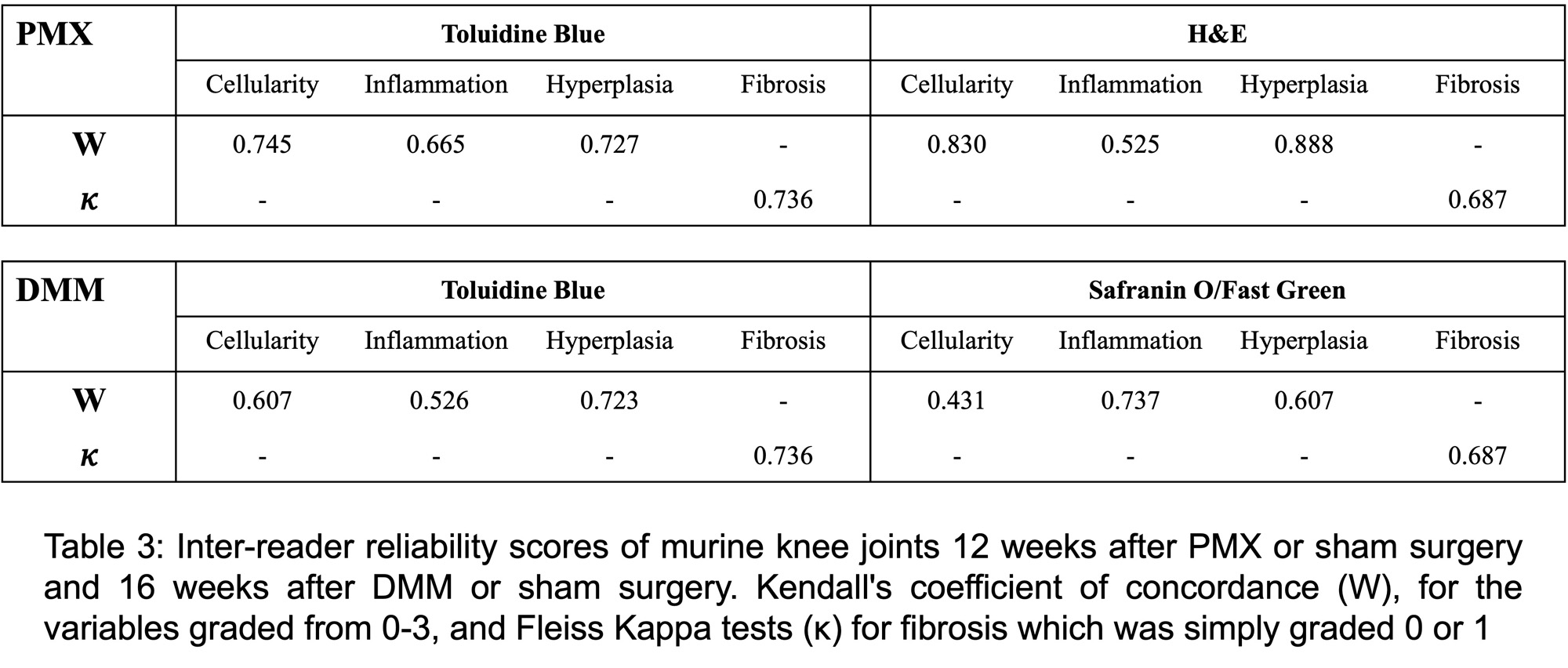
Methods: Coronal sections from murine knee joints as part of other studies were used as follows: 1. Hematoxylin and Eosin (H&E) or Toluidine Blue (T-Blue) stained sections from mice 6 weeks after destabilization of medial meniscus (DMM) or sham surgery and age matched naïve controls (n=5/group), 2. Safranin O (Saf-O) sections from mice 16 weeks after DMM or sham surgery (n=5-6/group), 3. T-Blue sections from mice 16 weeks after DMM or sham surgery (n=10/group), 4. T-Blue sections from 12 weeks after partial meniscectomy (PMX) or sham surgery (n=10/group), 5. H&E sections from 12 weeks after PMX or sham surgery (n=5/group). Four blinded readers graded four pathological features (hyperplasia, cellularity, inflammation, and fibrosis) at four anatomic locations (lateral and medial femoral gutters, and lateral and medial tibial gutters) using midjoint sections. Inter-reader reliability tests (IRR) (Kendall’s coefficient of concordance (W) for hyperplasia, inflammation, and cellularity; Fleiss Kappa test (κ) for fibrosis) were performed to assess agreement between readers.
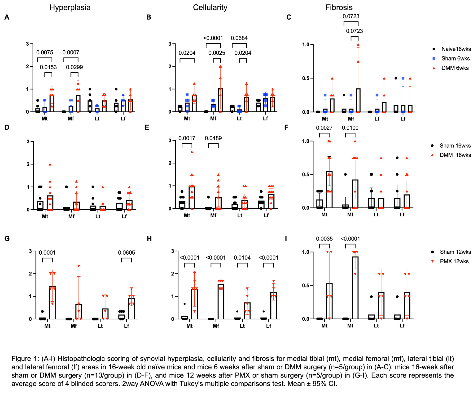
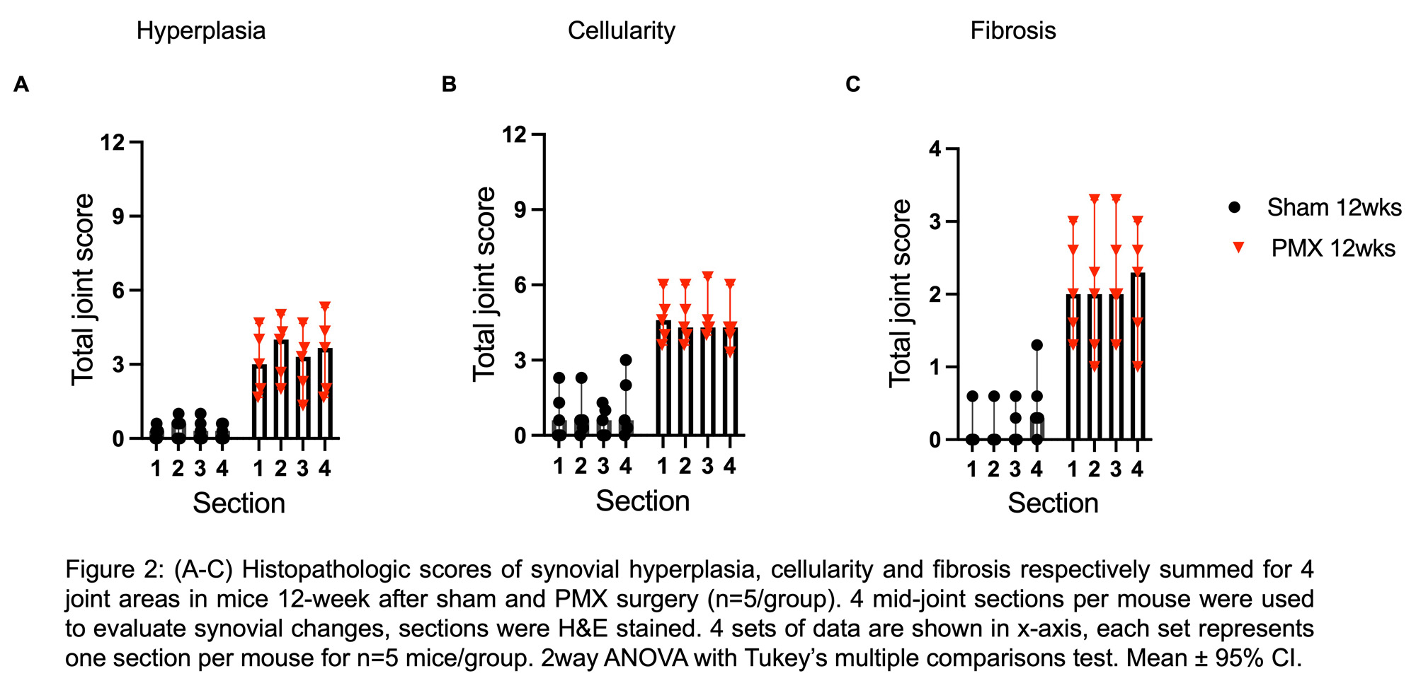
Results: IRR measures (Table 1) demonstrate that there was acceptable to very good agreement between raters. Inflammation scores were excluded because of poorer concordance between readers and overlap with the cellularity assessment. Increased hyperplasia and cellularity and a trend of increased fibrosis were observed in the medial tibial and medial femoral gutters compared to controls 6wks after DMM (Fig 1A-C). At 16-weeks post-DMM, significant increase in cellularity and fibrosis were observed in the medial side with no significant hyperplasia (Fig 1D-F). No significant lateral changes were observed in any of DMM groups. At 12 weeks after PMX, a significant increase in hyperplasia, cellularity and fibrosis were observed in the medial gutters compared to sham mice (Fig 1G-I). Increased cellularity and hyperplasia were evident in the lateral gutters of PMX mice. Additionally, we assessed 4 sections/knee from PMX mice and found that synovial changes were consistent from section to section (Fig 2). Finally, we assessed the effect of different stains on IRR (Table 1). Higher reliability was observed using H&E and T-blue stains.
Conclusion: Based on our findings, we suggest evaluating 3 pathological features at 4 anatomic areas of the joint to assess synovial pathology in murine models of OA. A minimum of 3 readers are recommended, for which the IRR should be determined. A single midjoint section is likely sufficient for analysis, and a careful choice of stain should be considered. It is important to note that these recommendations are considered an initial step to allow comparison across labs and models, and to be used to guide more detailed subsequent analyses.
To cite this abstract in AMA style:
Obeidat A, Kim S, Hu B, Li J, Ishihara S, Xiao R, Miller R, Malfait A, Scanzello C. Recommendations for a Standardized Approach to Histopathologic Evaluation of Synovial Membrane in Murine Models of Experimental Osteoarthritis [abstract]. Arthritis Rheumatol. 2023; 75 (suppl 9). https://acrabstracts.org/abstract/recommendations-for-a-standardized-approach-to-histopathologic-evaluation-of-synovial-membrane-in-murine-models-of-experimental-osteoarthritis/. Accessed .« Back to ACR Convergence 2023
ACR Meeting Abstracts - https://acrabstracts.org/abstract/recommendations-for-a-standardized-approach-to-histopathologic-evaluation-of-synovial-membrane-in-murine-models-of-experimental-osteoarthritis/


