ACR Meeting Abstracts

ACR Meeting Abstracts

  • Meetings
    • ACR Convergence 2024
    • ACR Convergence 2023
    • 2023 ACR/ARP PRSYM
    • ACR Convergence 2022
    • ACR Convergence 2021
    • ACR Convergence 2020
    • 2020 ACR/ARP PRSYM
    • 2019 ACR/ARP Annual Meeting
    • 2018-2009 Meetings
    • Download Abstracts
  • Keyword Index
  • Advanced Search
  • Your Favorites
    • Favorites
    • Login
    • View and print all favorites
    • Clear all your favorites
  • ACR Meetings

Abstract Number: 0655

Proteomic Analysis of Histological Lesions Lupus Nephritis Identifies an Inflammatory Signature of Fibrous Crescents

Alessandra Ida Celia1, Jeffery Hodgin2, Avi Rosenberg3, Laurence S Magder4, Jill Buyon5, Betty Diamond6, Judith James7, William Apruzzese8, Paride Fenaroli9, Derek Fine1, Jose Monroy-Trujillo1, Mohamed G. Atta1, Peter izmirly10, Michael Belmont5, Anne Davidson6, Daniel W. Goldman11, the Accelerating Medicines Partnership (AMP) RA/SLE12, Michelle Petri11 and Andrea Fava1, 1Johns Hopkins University, Baltimore, MD, 2University of Michigan, Ann Arbor, MI, 3Johns Hopkins University, Ballwin, MO, 4University of Maryland, Department of Epidemiology and Public Health, Baltimore, MD, 5NYU Grossman School of Medicine, New York, NY, 6Feinstein Institutes for Medical Research, Manhasset, NY, 7Oklahoma Medical Research Foundation, Oklahoma City, OK, 8Brigham and Women's Hospital, Boston, MA, 9Università degli Studi di Parma, Parma, Emilia-Romagna, Italy, 10NYU Long Island School of Medicine, New York, NY, 11Johns Hopkins University School of Medicine, Division of Rheumatology, Baltimore, MD, 12Multiple Insitutions

Meeting: ACR Convergence 2022

Keywords: Biomarkers, chemokines, Lupus nephritis, Renal, Systemic lupus erythematosus (SLE)

  • Tweet
  • Click to email a link to a friend (Opens in new window) Email
  • Click to print (Opens in new window) Print
Session Information

Date: Sunday, November 13, 2022

Title: SLE – Etiology and Pathogenesis Poster

Session Type: Poster Session B

Session Time: 9:00AM-10:30AM

Background/Purpose: We employed urine proteomics to define the molecular signatures associated with the histological features quantified by the NIH activity and chronicity indices.

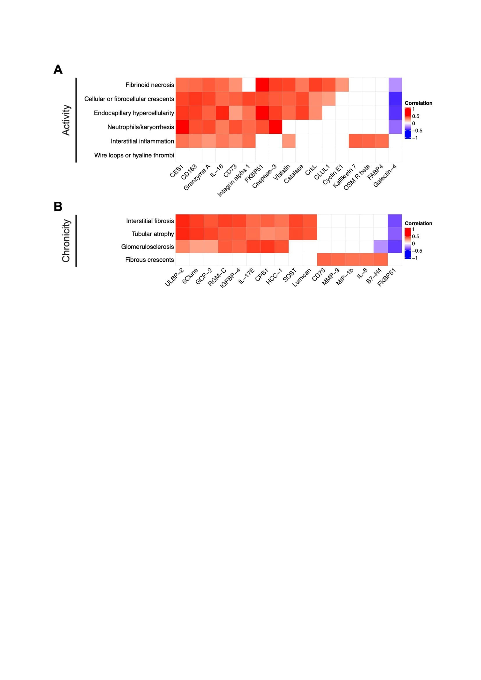
Methods: Glomerular and interstitial lesions in lupus nephritis were quantified (scored 0-3) based on the revised 2018 International Society of Nephrology/Renal Pathology Society (ISN/RPS) classification for lupus nephritis and the modified NIH scoring system by a central renal pathologist (JH). Urinary proteins (1200 biomarkers, RayBiotech Kiloplex) were quantified in urine samples collected on the day of (73%) or within 3 weeks of (27%) the diagnostic kidney biopsy. Proteomic signatures of each lesion were defined based on Spearman correlations of each urine protein with each pathologic lesion.

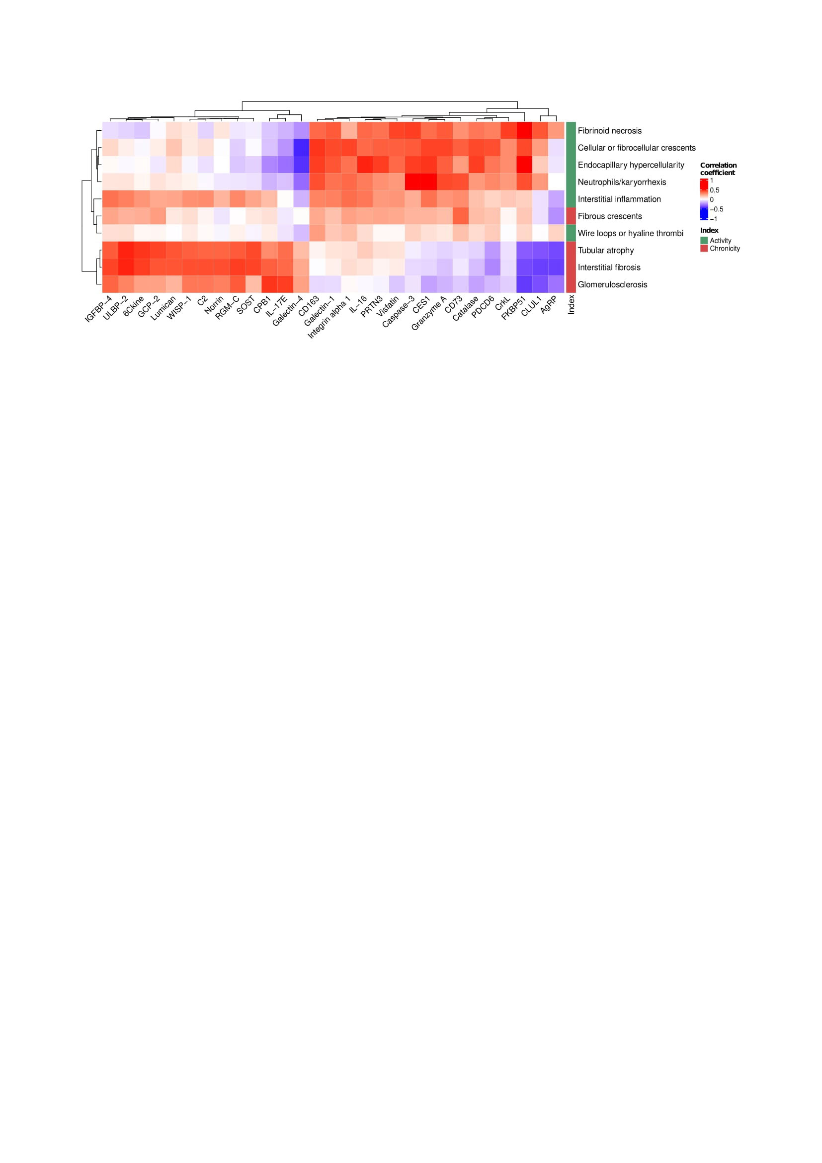
Results: Ninety-one biopsies were included: 32 (35%) with pure proliferative LN, 33 (36%) with pure membranous LN, and 26 (29%) with mixed LN. The 5 most correlated urinary proteins and each pathologic feature are summarized in Figure 1. Most lesions in the activity or chronicity indices shared a similar signature within their respective index. In contrast, fibrous crescents displayed an inflammatory signature (CD73, MMP9, MIP1b, and IL-8) despite being part of the NIH chronicity index. Hierarchical clustering based on proteomic signatures revealed that fibrous crescents were more similar to activity-related lesions (Figure 2). Interstitial inflammation (activity) was correlated with biomarkers associated with both active and chronic lesions.

Conclusion: Although fibrous crescents are considered inactive lesions that follow crescentic glomerulonephritis, urine proteomics revealed inflammatory activity associated with fibrous crescents. Several cell types such macrophages, fibroblasts, neutrophils, lymphocytes, and epithelial cells are critical in the formation of crescents. Higher levels of CD73, IL-8 and MMP9 indicate the presence of an inflammatory response involved in glomerular remodeling after extra-capillary proliferation that have an important role in kidney damage. The presence of fibrous crescents in kidney biopsies may indicate ongoing potentially treatable inflammation. Interstitial inflammation, which is linked to worse clinical outcomes, showed a distinct proteomic signature combining both activity and chronicity. A better understanding of the pathophysiology of processes including fibrous crescents and interstitial inflammation is needed to tailor treatment of these pathways leading to chronic damage.

Supporting image 1

Figure 1. Proteomic signatures of LN histological lesions. Heatmap displaying Spearman correlations of urinary proteins and any single subitem of the NIH activity (A) and chronicity (B) indices. For each lesion, the 5 most correlated proteins are displayed. Empty squares indicate a false discovery (FDR) rate > 0.25.

Supporting image 2

Figure 2. Fibrous crescents cluster with LN activity lesions. Hierarchical clustering based on the correlations of each histological lesion and urinary proteins. All proteins with a strict statistically significant correlation (FDR < 0.01) with at least one histological lesion were included.


Disclosures: A. Celia, None; J. Hodgin, None; A. Rosenberg, None; L. Magder, None; J. Buyon, Equillium, GlaxoSmithKlein(GSK), L and M Healthcare Communications, Janssen, Boomcom, Merck/MSD; B. Diamond, None; J. James, Bristol-Myers Squibb(BMS), AstraZeneca, Novartis, Progentec Biosciences; W. Apruzzese, None; P. Fenaroli, None; D. Fine, None; J. Monroy-Trujillo, None; M. Atta, Kira, Horizon, Bristol-Myers Squibb(BMS), REATA, Morphosys AG, Sentien; P. izmirly, None; M. Belmont, None; A. Davidson, None; D. Goldman, None; t. (AMP) RA/SLE, None; M. Petri, Exagen, AstraZeneca, Alexion, Amgen, AnaptysBio, Argenx, Aurinia, Biogen, Caribou Biosciences, CVS Health, EMD Serono, Eli Lilly, Emergent Biosolutions, GlaxoSmithKline (GSK), IQVIA, Janssen, Kira Pharmaceuticals, MedShr, Sanofi, SinoMab, Thermofisher, BPR Scientific Advisory Committee; A. Fava, Sanofi.

To cite this abstract in AMA style:

Celia A, Hodgin J, Rosenberg A, Magder L, Buyon J, Diamond B, James J, Apruzzese W, Fenaroli P, Fine D, Monroy-Trujillo J, Atta M, izmirly P, Belmont M, Davidson A, Goldman D, (AMP) RA/SLE t, Petri M, Fava A. Proteomic Analysis of Histological Lesions Lupus Nephritis Identifies an Inflammatory Signature of Fibrous Crescents [abstract]. Arthritis Rheumatol. 2022; 74 (suppl 9). https://acrabstracts.org/abstract/proteomic-analysis-of-histological-lesions-lupus-nephritis-identifies-an-inflammatory-signature-of-fibrous-crescents/. Accessed .
  • Tweet
  • Click to email a link to a friend (Opens in new window) Email
  • Click to print (Opens in new window) Print

« Back to ACR Convergence 2022

ACR Meeting Abstracts - https://acrabstracts.org/abstract/proteomic-analysis-of-histological-lesions-lupus-nephritis-identifies-an-inflammatory-signature-of-fibrous-crescents/

Advanced Search

Your Favorites

You can save and print a list of your favorite abstracts during your browser session by clicking the “Favorite” button at the bottom of any abstract. View your favorites »

All abstracts accepted to ACR Convergence are under media embargo once the ACR has notified presenters of their abstract’s acceptance. They may be presented at other meetings or published as manuscripts after this time but should not be discussed in non-scholarly venues or outlets. The following embargo policies are strictly enforced by the ACR.

Accepted abstracts are made available to the public online in advance of the meeting and are published in a special online supplement of our scientific journal, Arthritis & Rheumatology. Information contained in those abstracts may not be released until the abstracts appear online. In an exception to the media embargo, academic institutions, private organizations, and companies with products whose value may be influenced by information contained in an abstract may issue a press release to coincide with the availability of an ACR abstract on the ACR website. However, the ACR continues to require that information that goes beyond that contained in the abstract (e.g., discussion of the abstract done as part of editorial news coverage) is under media embargo until 10:00 AM ET on November 14, 2024. Journalists with access to embargoed information cannot release articles or editorial news coverage before this time. Editorial news coverage is considered original articles/videos developed by employed journalists to report facts, commentary, and subject matter expert quotes in a narrative form using a variety of sources (e.g., research, announcements, press releases, events, etc.).

Violation of this policy may result in the abstract being withdrawn from the meeting and other measures deemed appropriate. Authors are responsible for notifying colleagues, institutions, communications firms, and all other stakeholders related to the development or promotion of the abstract about this policy. If you have questions about the ACR abstract embargo policy, please contact ACR abstracts staff at [email protected].

Wiley

  • Online Journal
  • Privacy Policy
  • Permissions Policies
  • Cookie Preferences

© Copyright 2025 American College of Rheumatology