Session Information
Session Type: Poster Session A
Session Time: 8:30AM-10:30AM
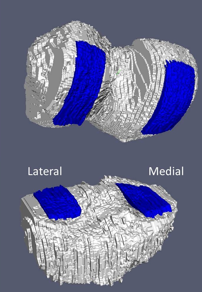
Background/Purpose: Semi-automated Local Area Cartilage Segmentation (LACS) software uses two robust coordinate systems to measure cartilage change in focused regions in the femur and tibia. (Figure 1) LACS also offers a topological method to assess change in cartilage volume through the use of responsiveness heat-maps. The purpose of this study was to provide a descriptive assessment of cartilage volume change in the medial femur (MF) and medial tibia (MT) for different (Kellgren-Lawrence) KL grades with the hypothesis that there will be evidence for different patterns of changes in cartilage volume across KL grades 1, 2 and 3.
Methods: Cartilage volume was measured in a central weight-bearing portion of the MF and MT using the LACS method at the baseline (BL) and 48 month visits of 670 participants from the Osteoarthritis Initiative (OAI), a longitudinal observational cohort study. Participants with at least one knee KL 1, 2, or 3 on centrally read baseline posterior-anterior weight-bearing knee radiographs were selected. Participants with a prosthetic knee or KL4 knee at baseline were excluded. One knee per participant was selected, based on maximum KL grade or randomly, if both knees had the same KL grade. Cartilage volume change was calculated as standardized response mean (SRM) for both plates separately. LACS SRM was estimated over grids in the sample and displayed in the form of responsiveness heat-maps, showing topographical change as SRM values over each cartilage plate region. (i.e., blue sub-regions in Figure 1) The mathematical nature of the LACS method permits any grid location to be determined consistently.
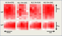
Results: At baseline, the participants had a mean age of 61.3 years, a mean BMI of 28.9, and were 41.5% male. The baseline distribution of KL grades was: KL1: 23.9% (N=160), KL2: 47.1% (N=316), KL3: 29.0% (N= 194). SRM values are shown in Table 1. These data indicate that the cartilage volume change is similar between KL1 and KL2 with a substantial increase for KL3. The responsiveness heat-maps are shown in Figure 2. We observed different patterns of change across the heat-maps. With increasing KL grade from 1 to 3, there appeared to be greater amount of change in the lateral portion of both the MF and MT compartments with the MT and MF having similar patterns of change.
Conclusion: As expected, loss of cartilage volume is increased in KL3 knees. But the heat-maps suggest there is more cartilage loss in the inner (lateral) portion of the knee in patients with more advanced OA as seen by a modestly higher intensity in the right-hand side of each heatmap with increasing KL grade. The variation in patterns of change in cartilage volume by KL grade demonstrate that the heat-map approach may offer a way to characterize different disease-related patterns of knee osteoarthritis progression. Additional studies will be necessary to better understand the role of responsiveness heat-maps. In the future, this work will be extended to the lateral compartment, and differences by gender, BMI, age, knee alignment, and other clinical variables will be assessed.
To cite this abstract in AMA style:
Amesbury R, Ragati Haghi H, Laffaye T, Stein R, Ashbeck E, Kwoh C, Mathiessen A, Duryea J. Distribution of Medial Femur and Tibia Cartilage Volume Change over 48 Months on MRI: Comparison Among Kellgren-Lawrence Grades [abstract]. Arthritis Rheumatol. 2021; 73 (suppl 9). https://acrabstracts.org/abstract/distribution-of-medial-femur-and-tibia-cartilage-volume-change-over-48-months-on-mri-comparison-among-kellgren-lawrence-grades/. Accessed .« Back to ACR Convergence 2021
ACR Meeting Abstracts - https://acrabstracts.org/abstract/distribution-of-medial-femur-and-tibia-cartilage-volume-change-over-48-months-on-mri-comparison-among-kellgren-lawrence-grades/


