Session Information
Session Type: Poster Session D
Session Time: 9:00AM-11:00AM
Background/Purpose: Ectopic soft tissue deposition of hydroxyapatite (calcinosis) is a frequent and morbid complication of dermatomyositis and scleroderma with no known effective pharmacologic treatment. 18F-NaF PET/CT detects active hydroxyapatite deposition in benign and malignant bone disorders and may identify subclinical and actively calcifying lesions in scleroderma- and dermatomyositis-related calcinosis.
Methods: In this pilot study, we enrolled three adults with dermatomyositis and three adults with scleroderma, all with new calcinosis deposits within the past six months. Each participant underwent 18F-NaF PET/CT as well as a clinical examination for assessment of calcinosis. We recorded the number, locations, and standardized uptake values (SUVs) of deposits for each 18F-NaF PET/CT.
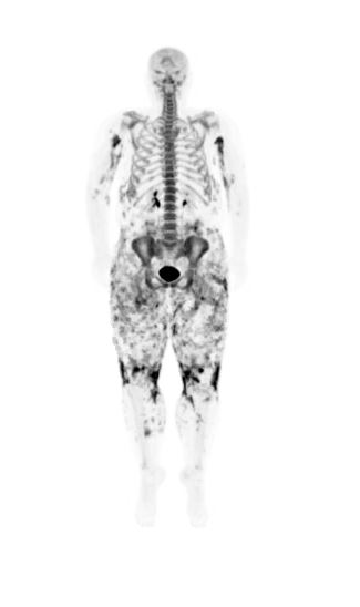
Results: 18F-NaF PET/CT detected heterogeneous uptake in calcinosis deposits appearing homogeneous on CT. 18F-NaF PET/CT also detected calcium uptake where there was no visible calcification on CT. We noted increased uptake of 18F-NaF in the axial skeleton of one patient with multiple actively calcifying deposits, consistent with increased systemic bone turnover. Some joints also demonstrated increased 18F-NaF uptake, consistent with arthritic remodeling.
Conclusion: 18F-NaF uptake on PET/CT is heterogeneous, even in radiographically homogeneous deposits, and identifies actively calcifying and subclinical deposits not apparent on CT. 18F-NaF uptake on PET/CT may be an early and sensitive marker of disease activity in calcinosis due to scleroderma and dermatomyositis.
 Axial CT image in bone windows (A) demonstrates calcinosis involving the quadriceps and patellar tendon complex overlying the left patella. Axial 18F-NaF PET/CT images (B and C) demonstrate mild activity along this calcified complex (blue arrowheads). However, there is focal PET signal activity located along the semimebranosus muscle without associated calcification on CT (red arrowheads). These findings may suggest increased sensitivity of 18F-NaF PET imaging in detecting pre-calcified disease involvement.
 Full-body coronal 18F-NaF PET image of Patient 6 demonstrating the distribution and intensity of 18F-NaF uptake. Note the intense activity in the upper arms and around the knees as well as widespread uptake in the thighs and pelvic girdle. There is also increased uptake in the axial skeleton, indicating increased systemic bone turnover
To cite this abstract in AMA style:
Richardson C, Javadi M, Shah A, Solnes L, Wigley F, Hummers L, Christopher-Stine L. 18F-NaF PET/CT Identifies Active Calcium Uptake in Calcinosis Due to Dermatomyositis and Scleroderma [abstract]. Arthritis Rheumatol. 2020; 72 (suppl 10). https://acrabstracts.org/abstract/18f-naf-pet-ct-identifies-active-calcium-uptake-in-calcinosis-due-to-dermatomyositis-and-scleroderma/. Accessed .« Back to ACR Convergence 2020
ACR Meeting Abstracts - https://acrabstracts.org/abstract/18f-naf-pet-ct-identifies-active-calcium-uptake-in-calcinosis-due-to-dermatomyositis-and-scleroderma/
