Session Information
Date: Sunday, November 8, 2015
Title: Rheumatoid Arthritis - Human Etiology and Pathogenesis Poster I
Session Type: ACR Poster Session A
Session Time: 9:00AM-11:00AM
Background/Purpose: Malondialdehyde-acetaldehyde
(MAA) adducts are expressed in synovial tissues in rheumatoid arthritis (RA). Post-translational
MAA modifications are pro-inflammatory, promoting robust anti-MAA antibody
responses that are correlated with markers of RA disease severity. Whether
anti-MAA antibodies are produced locally in affected joint tissues is not
known. If present in higher concentrations in synovial fluid (SF) than in
sera, this would support a potential pathogenic role of these antibodies in RA.
The objective of this study was to evaluate the expression of anti-MAA antibody
isotypes in paired SF/serum samples of patients with RA and OA to better
understand the relationship of circulating antibody with antibody expressed
locally in joints.
Methods: Paired serum/SF samples were
examined, drawn from 29 RA patients and 13 OA patients on the same day, banked,
and frozen at -700C until analysis. Anti-MAA antibody isotypes (IgA,
IgG, and IgM) were measured in arbitrary units (AU) using ELISA and examined as
means ± SEMs.
Paired t-tests were used to compare anti-MAA antibody concentrations in
SF vs. serum, while RA/OA comparisons were made separately for SF and serum
using a Student’s t-test.
Results:
Anti-MAA antibody
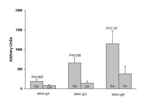
concentrations were numerically higher in RA-SF vs. OA-SF for IgA and IgM, a
difference that reached statistical significance for the IgG isotype (p=0.008)
(Figure 1). There were no significant RA-OA differences for any of the
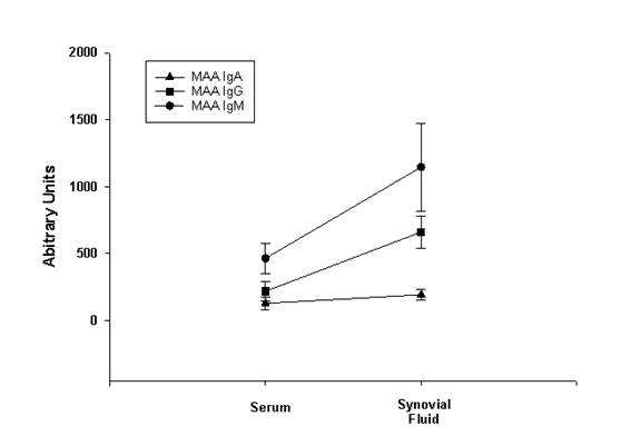
circulating anti-MAA antibody isotypes. In analyses of paired RA samples, IgG (663±121
vs. 219±76 AU, p<0.001) and IgM (1148±328 vs. 464±115 AU, p=0.033) anti-MAA
antibody isotypes were significantly higher in the SF vs. serum. IgA anti-MAA
antibody concentrations in RA SF did not differ significantly from paired sera
(Figure 2). There
were no differences in
anti-MAA antibody concentrations in paired SF/serum samples from OA patients.
Conclusion:
These results
complement a previous report demonstrating that MAA adducts are expressed in inflammatory
joint tissues in patients with RA. For the first time, these results show that
anti-MAA antibodies, particularly the IgM and IgG isotypes, are also present in
significantly higher concentrations in the joint compared to the circulation.
It is possible that anti-MAA antibody may be produced locally or alternatively
that these antibodies accumulate in joint tissues. Taken together, these
results suggest that anti-MAA antibody could play an important pathogenic role and
that its detection in SF could serve as an informative RA biomarker.
Figure 1:
Figure 2:
To cite this abstract in AMA style:
Rahman R, Thiele GM, Hollins A, Duryee MJ, Anderson D, Hamilton B, Michaud K, Klassen LW, Mikuls TR. Antibodies to Malondialdehyde-Acetaldehyde Adducts Are Highly Expressed in Rheumatoid Arthritis Synovial Fluid [abstract]. Arthritis Rheumatol. 2015; 67 (suppl 10). https://acrabstracts.org/abstract/antibodies-to-malondialdehyde-acetaldehyde-adducts-are-highly-expressed-in-rheumatoid-arthritis-synovial-fluid/. Accessed .« Back to 2015 ACR/ARHP Annual Meeting
ACR Meeting Abstracts - https://acrabstracts.org/abstract/antibodies-to-malondialdehyde-acetaldehyde-adducts-are-highly-expressed-in-rheumatoid-arthritis-synovial-fluid/


