Session Information
Session Type: Poster Session C
Session Time: 10:30AM-12:30PM
Background/Purpose: Chronic nonbacterial osteomyelitis (CNO) is an autoinflammatory bone disorder characterized by multifocal, sterile bone inflammation in childhood. Whole-body magnetic resonance imaging (WB-MRI) allows for a comprehensive assessment of skeletal involvement in CNO, which can enhance our understanding of the disorder and its implication for child wellbeing. Our objective was to provide a novel perspective of CNO, leveraging WB-MRI methods linked with clinical records to identify the demographic, clinical, and imaging characteristics of the children receiving care at a tertiary pediatric center.
Methods: We conducted a retrospective review of the pediatric patients diagnosed with CNO over a 10-year period (n=76). To be included children had to have a diagnosis of CNO confirmed by a pediatric rheumatologist, be less than 18 years old at time of diagnosis, and had a WB-MRI within 12 months of symptom onset. Demographic data, clinical features, extraosseous involvement, diagnostic and treatment modalities, and lesion distribution were ascertained by review of the electronic medical record. Lesion counts and locations were extracted from MRI reports and were validated by a musculoskeletal radiologist.
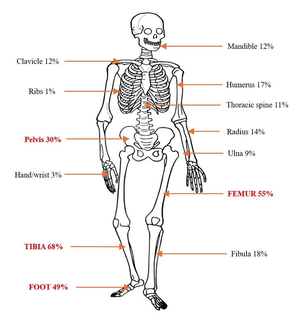
Results: Among the 76 patients (55% female), the median age at symptom onset was 10.2 years (Inter quartile range 7.7–12.8), and the median age at diagnosis was 10.7 years (IQR 8.2-13.1). WB-MRI was performed within a median 2.2 months after onset (IQR 0.5-5.7 months) and identified 410 lesions across the cohort. Asymptomatic lesions were common, highlighting the diagnostic value of WB-MRI. The median number of lesions per patient was 4 (range 1-23). The tibia (68%), the femur (55%) and the foot (49%) were the most frequently involved sites. Thoracic vertebral lesions were seen in 8 children. The mandible was involved in 9 children; interestingly 5 patients (56%) had extra-mandibular lesions. Extraosseous features including arthritis, psoriasis, and inflammatory bowel disease were seen only in a small subset of patients. HLA-B27 was positive in 16% of those tested. NSAIDs were the most used medication (93%), followed by TNF inhibitors (38%) and DMARDs (27%). At the last visit 19% were described by their pediatric rheumatologist as being in remission.
Conclusion: This is one of the largest single center cohorts of CNO with WB-MRI evaluation. Among children with CNO attending a tertiary care clinic, WB-MRI revealed a high burden of multifocal disease, often with clinically asymptomatic lesions. Confirming other published reports, CNO was more common in girls and onset age was around 10. Tibia, femur and the bones of the feet were frequently affected comparable to several multicenter CNO cohorts. Mandibular lesions were more frequent than in other reported cohorts. The diversity of lesion sites underscores the importance of WB-MRI in comprehensive evaluation. These findings support routine WB-MRI in the diagnostic work-up and monitoring of pediatric CNO.
 Characteristics of the CNO cohort
.jpg) Characteristics of skeletal lesions in patients with CNO
Characteristics of skeletal lesions in patients with CNO
.jpg) Distribution of lesions in patients with CNO
Distribution of lesions in patients with CNO
To cite this abstract in AMA style:
Rodriguez M, Davenport Munoz M, Braithwaite K, Ponder L, Rouster-Stevens K, Argeseanu Cunningham S, Prahalad S. Whole-Body MRI Findings in a Cohort of Children with Chronic Nonbacterial Osteomyelitis [abstract]. Arthritis Rheumatol. 2025; 77 (suppl 9). https://acrabstracts.org/abstract/whole-body-mri-findings-in-a-cohort-of-children-with-chronic-nonbacterial-osteomyelitis/. Accessed .« Back to ACR Convergence 2025
ACR Meeting Abstracts - https://acrabstracts.org/abstract/whole-body-mri-findings-in-a-cohort-of-children-with-chronic-nonbacterial-osteomyelitis/