ACR Meeting Abstracts

ACR Meeting Abstracts

  • Meetings
    • ACR Convergence 2024
    • ACR Convergence 2023
    • 2023 ACR/ARP PRSYM
    • ACR Convergence 2022
    • ACR Convergence 2021
    • ACR Convergence 2020
    • 2020 ACR/ARP PRSYM
    • 2019 ACR/ARP Annual Meeting
    • 2018-2009 Meetings
    • Download Abstracts
  • Keyword Index
  • Advanced Search
  • Your Favorites
    • Favorites
    • Login
    • View and print all favorites
    • Clear all your favorites
  • ACR Meetings

Abstract Number: 1901

What Does Radiographic Minimum JSW Actually Measure? Comparison of Radiographic and MRI Measures Using Data from the OAI

Alan Brett1, Michael Bowes2 and Philip G Conaghan3, 1Stryker, Austin, TX, 2Stryker, Manchester, United Kingdom, 3University of Leeds, Leeds, United Kingdom

Meeting: ACR Convergence 2022

Keywords: Biomarkers, clinical trial, Magnetic resonance imaging (MRI), Osteoarthritis, X-ray

  • Tweet
  • Click to email a link to a friend (Opens in new window) Email
  • Click to print (Opens in new window) Print
Session Information

Date: Monday, November 14, 2022

Title: Osteoarthritis – Clinical Poster

Session Type: Poster Session D

Session Time: 1:00PM-3:00PM

Background/Purpose: Change in radiographic joint space width (JSW) is used as a surrogate for cartilage loss and recognized by regulatory bodies as a measure of structural modification in DMOAD trials. However, both a reduction in cartilage thickness and meniscal integrity are inferred from a reduction in JSW. JSW measurement may be measured at any position in either the medial or lateral compartment. Often, the minimum JSW (minJSW) is measured because it is easily defined. However, the position of this minimum in the compartment can change between timepoints in a longitudinal study. Although the use of software can more reproducibly measure JSW at an arbitrary fixed position with greater sensitivity to change [1], the minJSW has persisted as a structural quantitative OA measure. More recently, minJSW has been used in the definition of Rapidly Progressing OA (RPOA1) in anti-NGF pharmaceutical trials.

We aimed to determine (i) the most common position for radiographic minJSW measurement in the medial compartment; (ii) the relative contributions of the average meniscal height and cartilage thickness at that position measured from 3D MR images; and (iii) how this compares to a fixed position medial JSW at the x=0.225 position [1] on a unit scale across the femur.

Methods: We used all 5,563 baseline knee images from the OAI that had minJSW measures to determine the median position of minJSW in the medial compartment for various Kellgren-Lawrence Grades (KLG) and OARSI JSN Grades. We performed careful manual segmentation of the meniscus and cartilage on a convenience set of 179 baseline OAI MRI DESS-we images, randomly chosen to provide representative samples of KL0/1, KL2 and KL3 grades. The medial tibial plateau of a mean 3D statistical shape model (SSM) of the knee was divided into 0.025-width anterior-posterior strips. These strips were projected from the mean SSM to each of the 179 3D knee examples and the relative contributions of meniscal height and cartilage to the total femur-tibia inter-bone distance were computed along each strip. A nearest strip was chosen to correspond with the median position of minJSW and compared with the strip at x=0.225.

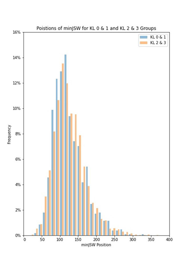
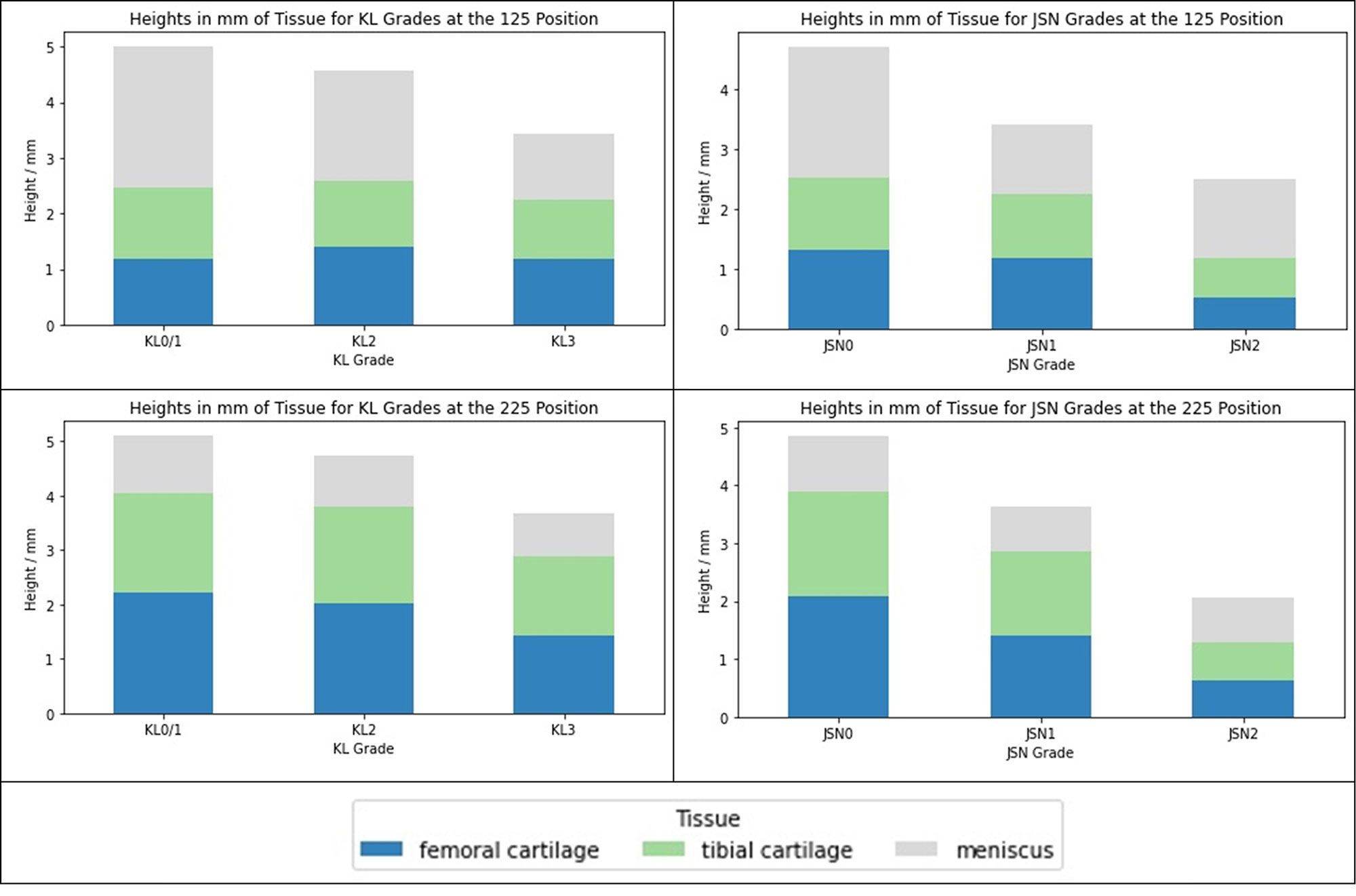
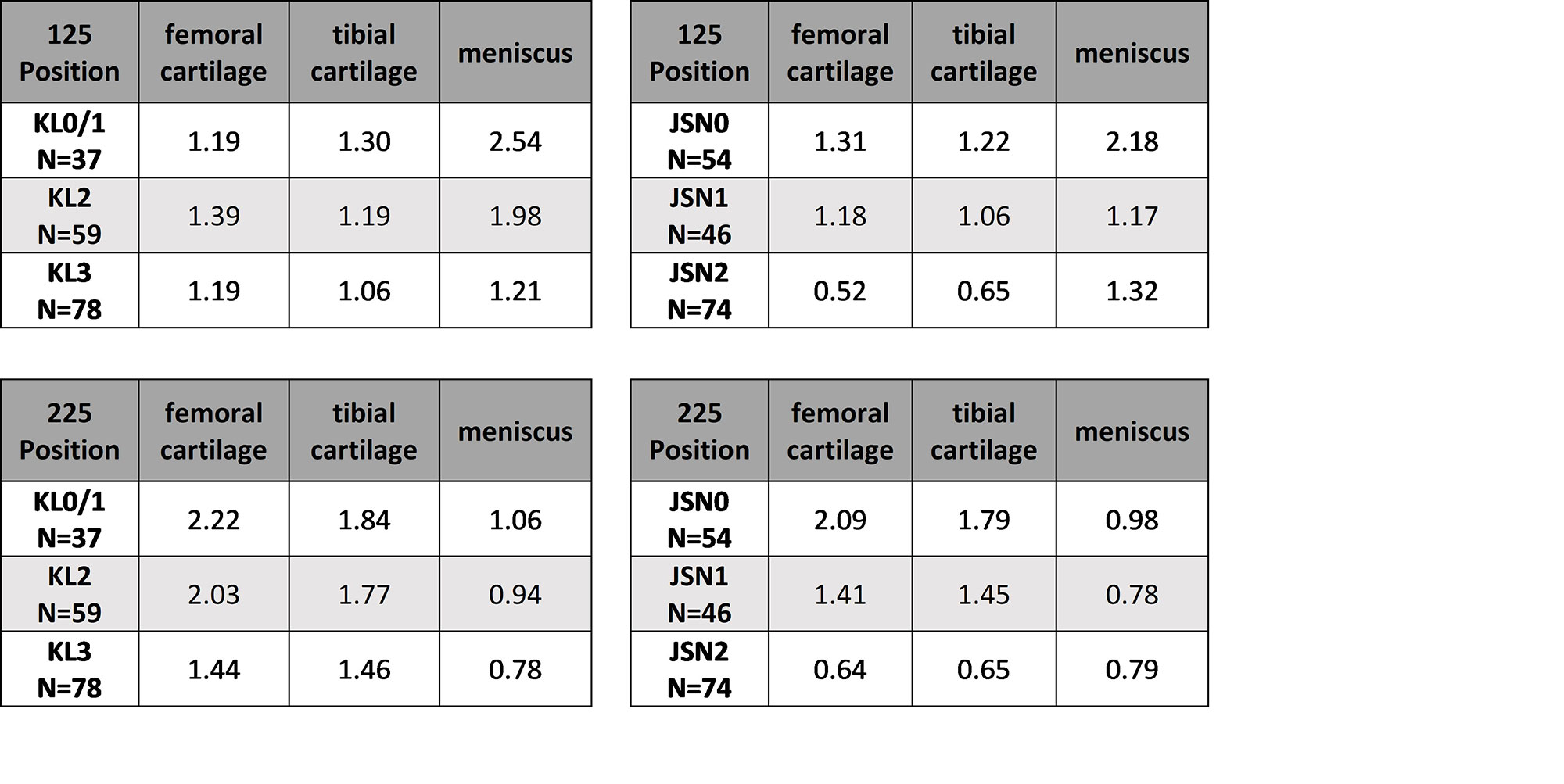
Results: Median medial compartment minJSW was found at the 0.119 position, this was consistent between KL0/1 and KL2/3 groups (Figure 1). The strip centered on x=0.125 was chosen to represent this position. Relative contributions of meniscus height and cartilage thickness to the inter-bone distance for the 0.125 and 0.225 positions are shown in Figure 2 and Table 1.

Conclusion: At the minJSW position (x=0.125), changes in meniscal height account for almost all of the differences between KLG and JSN groups, though cartilage does decrease significantly in the JSN2 group. At the 0.225 position, there is little difference in meniscal height at any stage of the disease, cartilage changes dominate the differences between all groups. While minJSW may be a useful measure of OA disease state, it is primarily a surrogate of meniscal and not cartilage changes, in contrast to the fixed position JSW measurements in the centre of the medial compartment, which better represents changes in the cartilage.

[1] Wirth, W. et al (2013). O&C 21(1), 117–125.

Supporting image 1

Figure 1: Histogram of positions of minJSW for KL0/1 and KL2/3 groups

Supporting image 2

Figure 2: Meniscal height, femoral cartilage thickness and tibial cartilage thickness in mm for different KLG and OARSI JSN at x=0.125 and x=0.225 positions.

Supporting image 3

Table 1: Meniscal height, femoral cartilage thickness and tibial cartilage thickness in mm for different KLG and OARSI JSN at x=0.125 and x=0.225 positions.


Disclosures: A. Brett, Stryker; M. Bowes, Stryker; P. Conaghan, AbbVie/Abbott, AstraZeneca, Bristol-Myers Squibb(BMS), Eli Lilly, Galapagos, GlaxoSmithKlein(GSK), Grunenthal, Merck/MSD, Novartis, Pfizer, Stryker, UCB, Amgen.

To cite this abstract in AMA style:

Brett A, Bowes M, Conaghan P. What Does Radiographic Minimum JSW Actually Measure? Comparison of Radiographic and MRI Measures Using Data from the OAI [abstract]. Arthritis Rheumatol. 2022; 74 (suppl 9). https://acrabstracts.org/abstract/what-does-radiographic-minimum-jsw-actually-measure-comparison-of-radiographic-and-mri-measures-using-data-from-the-oai/. Accessed .
  • Tweet
  • Click to email a link to a friend (Opens in new window) Email
  • Click to print (Opens in new window) Print

« Back to ACR Convergence 2022

ACR Meeting Abstracts - https://acrabstracts.org/abstract/what-does-radiographic-minimum-jsw-actually-measure-comparison-of-radiographic-and-mri-measures-using-data-from-the-oai/

Advanced Search

Your Favorites

You can save and print a list of your favorite abstracts during your browser session by clicking the “Favorite” button at the bottom of any abstract. View your favorites »

All abstracts accepted to ACR Convergence are under media embargo once the ACR has notified presenters of their abstract’s acceptance. They may be presented at other meetings or published as manuscripts after this time but should not be discussed in non-scholarly venues or outlets. The following embargo policies are strictly enforced by the ACR.

Accepted abstracts are made available to the public online in advance of the meeting and are published in a special online supplement of our scientific journal, Arthritis & Rheumatology. Information contained in those abstracts may not be released until the abstracts appear online. In an exception to the media embargo, academic institutions, private organizations, and companies with products whose value may be influenced by information contained in an abstract may issue a press release to coincide with the availability of an ACR abstract on the ACR website. However, the ACR continues to require that information that goes beyond that contained in the abstract (e.g., discussion of the abstract done as part of editorial news coverage) is under media embargo until 10:00 AM ET on November 14, 2024. Journalists with access to embargoed information cannot release articles or editorial news coverage before this time. Editorial news coverage is considered original articles/videos developed by employed journalists to report facts, commentary, and subject matter expert quotes in a narrative form using a variety of sources (e.g., research, announcements, press releases, events, etc.).

Violation of this policy may result in the abstract being withdrawn from the meeting and other measures deemed appropriate. Authors are responsible for notifying colleagues, institutions, communications firms, and all other stakeholders related to the development or promotion of the abstract about this policy. If you have questions about the ACR abstract embargo policy, please contact ACR abstracts staff at [email protected].

Wiley

  • Online Journal
  • Privacy Policy
  • Permissions Policies
  • Cookie Preferences

© Copyright 2025 American College of Rheumatology