Session Information
Session Type: Poster Session C
Session Time: 1:00PM-3:00PM
Background/Purpose: Superb microvascular imaging (SMI) is a novel technique that provides a more sensitive assessment of small vessels compared to color Doppler US (CDUS), by distinction of low-speed flow signals from motion artifacts. Superficial thrombophlebitis (STM) is a common manifestation in patients with Behçet syndrome and is thought to be associated with inflammation of the vessel wall rather than a procoagulant state. We aimed to assess STM lesions of patients with Behçet syndrome (BS), together with controls, using SMI to get a better understanding of these lesions.
Methods: We studied 45 BS (16 F/29 M, mean age: 40.0 ± 12.2) patients and 10 non-BS (6 F/4 M mean age: 45.0 ± 10.4) patients with nodular lesions on physical examination. B-mode US, CDUS and SMI were performed and recorded by the same radiologist (YK) and images were then evaluated by two radiologists (YK-AU). Both radiologists were blinded to the diagnoses and to each other’s assessments. Interobserver reliability was assessed by kappa statistic.
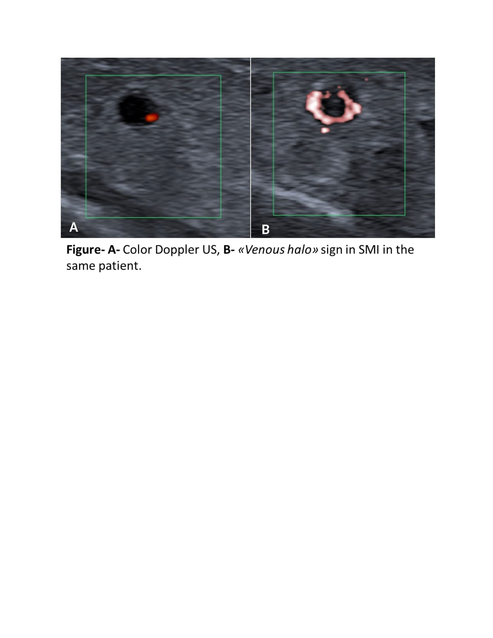
Results: The nodular lesions of 16 BS and 3 non-BS patients were diagnosed as STM. The diagnosis was phlebitis without thrombosis in 4 patients with BS and 1 patient with non-BS and erythema nodosum in the remaining 20 BS and 6 non-BS patients. A venous halo sign, meaning a halo shaped signal on the venous wall was detected with SMI in 13/16 (81%) BS patients with STM, 1 (25%) BS patient with phlebitis and none of the BS patients with erythema nodosum (Figure). Among the 3 non-BS patients with STM, 1 (33%) had venous halo sign. None of the other non-BS patients had a venous halo. The interobserver reliability was good (κ=0.96, p< 0.001) for detecting the venous halo sign.
Conclusion: A venous halo sign suggesting inflammation of the vessel wall was detected with SMI in the majority of BS patients with STM. This finding needs to be studied in different vascular lesions of a large number of BS patients together with controls, in order to understand its specificity for BS and its significance.
To cite this abstract in AMA style:
Kayadibi Y, Ozguler Y, Melikoglu M, Esatoglu S, Ustundag A, Kimyon U, Kalyoncu Ucar A, Adaletli I, Hatemi g. Venous Halo Sign Detected with Superb Microvascular Imaging in Behçet Syndrome [abstract]. Arthritis Rheumatol. 2022; 74 (suppl 9). https://acrabstracts.org/abstract/venous-halo-sign-detected-with-superb-microvascular-imaging-in-behcet-syndrome/. Accessed .« Back to ACR Convergence 2022
ACR Meeting Abstracts - https://acrabstracts.org/abstract/venous-halo-sign-detected-with-superb-microvascular-imaging-in-behcet-syndrome/

