Session Information
Date: Tuesday, November 9, 2021
Title: Metabolic & Crystal Arthropathies – Basic & Clinical Science Poster II (1565–1583)
Session Type: Poster Session D
Session Time: 8:30AM-10:30AM
Background/Purpose: Cardiovascular disease is a common comorbidity in people with gout. A hypothesized link between cardiovascular disease and gout is the deposition of monosodium urate (MSU) crystals in atherosclerotic plaque. Recent dual-energy CT (DECT) studies have reported that color-coded material consistent with MSU crystal deposition is present in calcified vessels of patients with gout. However, it is unclear whether these appearances represent true MSU crystal deposition or artifact during live imaging. The aim of this study was to examine whether MSU crystal deposition is present in the great vessels of cadaveric donors with gout by DECT or microscopic analysis.
Methods: Six cadaveric donors with a documented history of gout (two with visible tophi) were studied. Vascular tissue was dissected from the arch of the aorta through to the descending aorta at the level of the diaphragm, and included the proximal few centimeters of the brachiocephalic, common carotids, and subclavian arteries. Vascular tissue from each donor was scanned by DECT (SOMATOM Definition Flash, Siemens Medical, Erlangen, Germany) and analyzed for urate deposition on a Siemens workstation using proprietary gout software (syngo MMWP VE 36A 2009, Siemens Medical). After scanning, 100µl of phosphate buffered saline was injected into the vessel wall at ten standardized sites across each sample, and aspirated fluid was analyzed by polarizing light microscopy. Positive control tissue included the foot joints of cadaveric donors with tophaceous gout analyzed by DECT and microscopy using the same protocols, as well as synthesized MSU crystals as controls at the time of microscopic analysis.
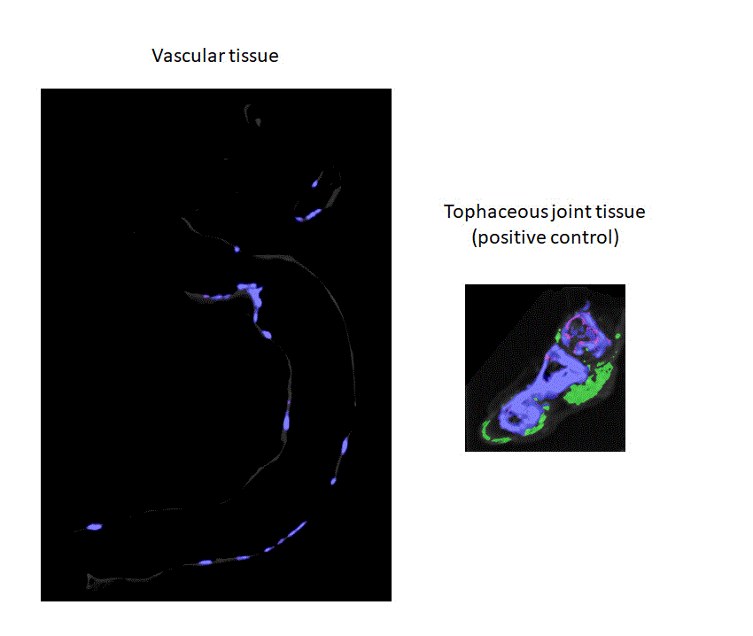
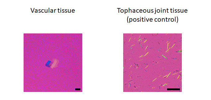
Results: DECT scanning of the cadaveric vascular tissue showed calcification in all samples, but no evidence of MSU crystal deposition (Figure 1). In contrast, urate deposition was clearly evident in the positive control tophaceous joint tissue by DECT. Microscopic analysis demonstrated plate-like cholesterol crystals from vascular tissue from five of the six donors (mean (SD) 3.2 (1.9) sites/donor, Figure 2). A single negatively birefringent needle shaped crystal was visualized from one of the 60 standardized sites analyzed, with no MSU crystals evident at any other site. In contrast, numerous negatively birefringent needle shaped crystals were present in aspirates from positive control tophaceous joint tissue.
Conclusion: This analysis has not demonstrated extensive MSU crystal deposition in vascular tissue of cadaveric donors with gout by DECT or polarizing light microscopy.
 Figure 1. Dual energy CT. Left panel. Representative DECT image of cadaveric vascular tissue demonstrating vascular calcification in blue, but no urate deposition (color-coded green). Right panel. Representative DECT image of positive control cadaveric tophaceous joint tissue demonstrating urate deposition (color-coded green) within tophaceous deposits. Nail artifact is also evident.
 Figure 2. Polarizing light microscopy. Left panel. Representative polarizing microscopy image from cadaveric vascular tissue demonstrating plate-like cholesterol crystals, but no needle-shaped negatively birefringent crystals. Scale bar 10µm. Right panel. Representative polarizing microscopy image from positive control cadaveric tophaceous joint tissue demonstrating numerous needle-shaped negatively birefringent crystals. Scale bar 10µm.
To cite this abstract in AMA style:
Dalbeth N, Alhilali M, Riordan P, Narang R, Chhana A, McGlashan S, Doyle A, ANDRES M. Vascular Monosodium Urate Crystal Deposition in Gout: A Dual-energy CT and Microscopy Study of Cadaveric Donors [abstract]. Arthritis Rheumatol. 2021; 73 (suppl 9). https://acrabstracts.org/abstract/vascular-monosodium-urate-crystal-deposition-in-gout-a-dual-energy-ct-and-microscopy-study-of-cadaveric-donors/. Accessed .« Back to ACR Convergence 2021
ACR Meeting Abstracts - https://acrabstracts.org/abstract/vascular-monosodium-urate-crystal-deposition-in-gout-a-dual-energy-ct-and-microscopy-study-of-cadaveric-donors/
