
Session Information
Session Type: Poster Session C
Session Time: 10:30AM-12:30PM
Background/Purpose: Medium vessel vasculitis (MVV), including polyarteritis nodosa (PAN), presents significant diagnostic challenges with patients frequently exhibiting non-specific systemic manifestations. While 18F-fluorodeoxyglucose PET/CT (PET/CT) is widely used for large vessel vasculitis, its application in MVV is unclear. A previous case series of 10 patients by Fagart et al. has shown that FDG-PET/CT can reveal characteristic abnormalities such as increased tracer uptake in muscle compartments and medium sized vessels. This study evaluates the diagnostic efficacy of PET CT in a cohort of patients with medium vessel vasculitis.
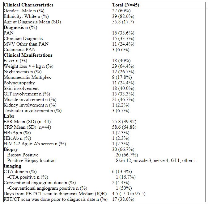
Methods: This retrospective study utilized data from Mayo Clinic’s records. We reviewed 369 charts and identified 45 patients diagnosed with MVV who underwent PET/CT scans for disease evaluation. Patients were categorized into four groups: PAN meeting classification criteria (n=16, 35.6%), clinician-diagnosed PAN (n=15, 33.3%), medium vessel vasculitis not diagnosed as PAN (n=11, 24.4%), and cutaneous PAN (n=3, 6.7%). Descriptive statistics are reported by group, and differences were tested using Kruskal-Wallis and Fisher’s exact tests.
All PET/CT scans were performed without intravenous contrast. They were considered positive if the vessel wall tracer activity was higher than the blood pool activity regardless of CT findings. Active myositis was defined as increased FDG accumulation higher than normal muscle background activity.
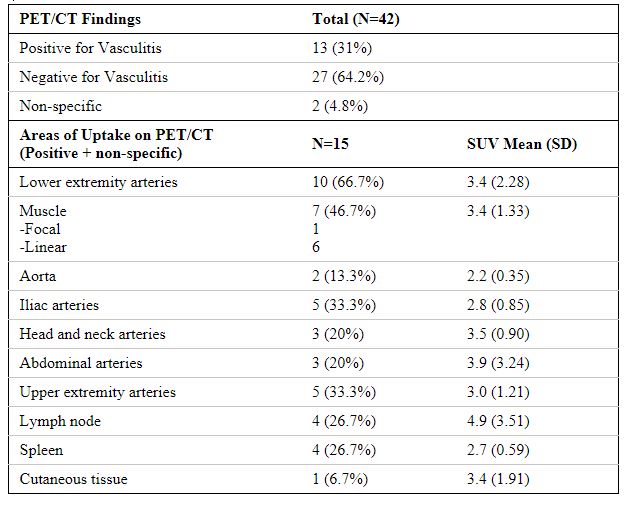
Results: Of the 45 patients, 27 (60%) were male and 39 (88.6%) were white, with a mean age at diagnosis of 55.8 years. Clinical manifestations included constitutional symptoms in 29 (64.4%), neurologic involvement in 19 (42.2%), and muscle involvement in 21 (46.7%). Biopsies were done in 30 patients and showed evidence of vasculitis in 20 (66.7%). Excluding those with cutaneous PAN, PET/CT scans were positive, i.e., showed evidence of FDG uptake in vessels, in 13 (31%) patients and negative in 27 (64.2%), 2 (4.8%) were non-specific (defined as muscle uptake only).
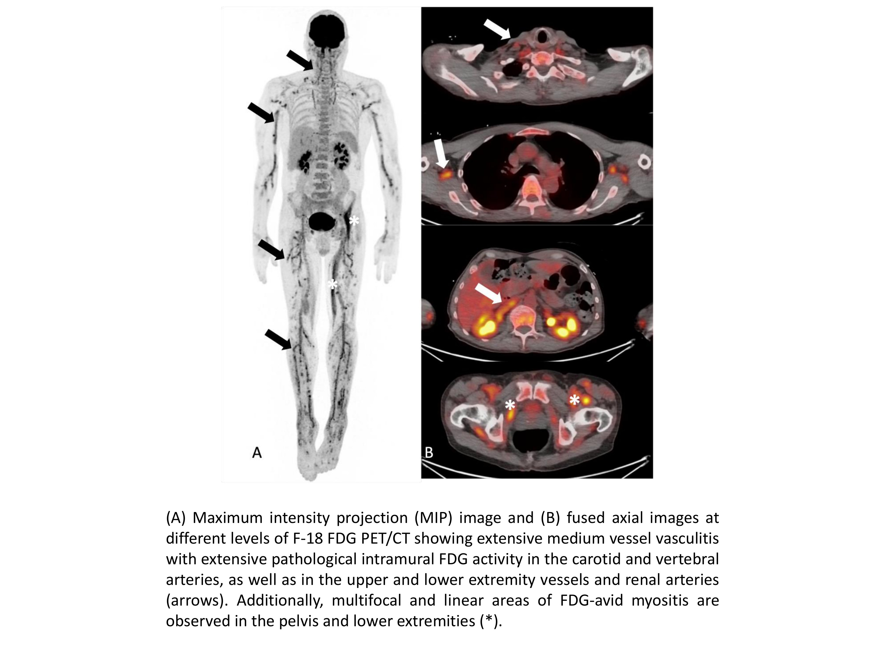
Common PET/CT findings included uptake in lower extremity vessels (10), muscles (7), and iliac arteries (5). The most common pattern of muscle uptake was linear (6/7). Location of uptake did not differ significantly between disease groups. Glucocorticoid use prior to PET/CT was common and reported in 19 (45.2%). Of those without prior glucocorticoid use 11/23 (47.8%) were positive versus 2/19 (10.5%) with prior use. Prior glucocorticoid use was inversely associated with a positive PET/CT (p=0.017).
Conclusion: PET/CT scans can show characteristic findings in MVV, with predominant involvement of lower extremity vessels and linear muscle uptake. However, there is limited sensitivity for diagnosing MVV and this can be further influenced by glucocorticoid use, highlighting the need for careful consideration of pre-scan treatment regimens. These findings suggest that PET/CT should be used with other diagnostic modalities for a comprehensive evaluation.
References:
1Fagart, A. et al. “Fluorodeoxyglucose positron emission tomography–computed tomography findings in a first series of 10 patients with polyarteritis nodosa.” Rheumatology 61.4 (2022): 1663-1668.
To cite this abstract in AMA style:
Eshak N, Yancey K, Martinez F, Mertz L, mead harvey c, Warrington K, Koster M, Sullivan M. Utility of 18 FDG-PET/CT Scans in the Diagnosis of Polyarteritis Nodosa and Other Medium Vessel Vasculitis [abstract]. Arthritis Rheumatol. 2024; 76 (suppl 9). https://acrabstracts.org/abstract/utility-of-18-fdg-pet-ct-scans-in-the-diagnosis-of-polyarteritis-nodosa-and-other-medium-vessel-vasculitis/. Accessed .« Back to ACR Convergence 2024
ACR Meeting Abstracts - https://acrabstracts.org/abstract/utility-of-18-fdg-pet-ct-scans-in-the-diagnosis-of-polyarteritis-nodosa-and-other-medium-vessel-vasculitis/