Session Information
Session Type: Poster Session A
Session Time: 8:30AM-10:30AM
Background/Purpose: Production of autoantibodies following pathogenic T and B cell interactions precede the development of RA. T follicular helper (Tfh) cells and T peripheral helper (Tph) cells are capable of providing B-cell help and both are increased in the peripheral blood in patients with seropositive RA. However, it is unknown whether these cell populations are expanded and/or altered in the peripheral blood prior to the onset of clinically apparent RA. Moreover, the contribution of these cells to the pathogenesis of RA may differ according to genetic and environmental backgrounds. Herein, we explored circulating Tph (cTph) cells and circulating Tfh (cTfh) cells from anti-citrullinated protein antibodies (ACPA)+ individuals both before and after development of RA in two ethnically distinct populations.
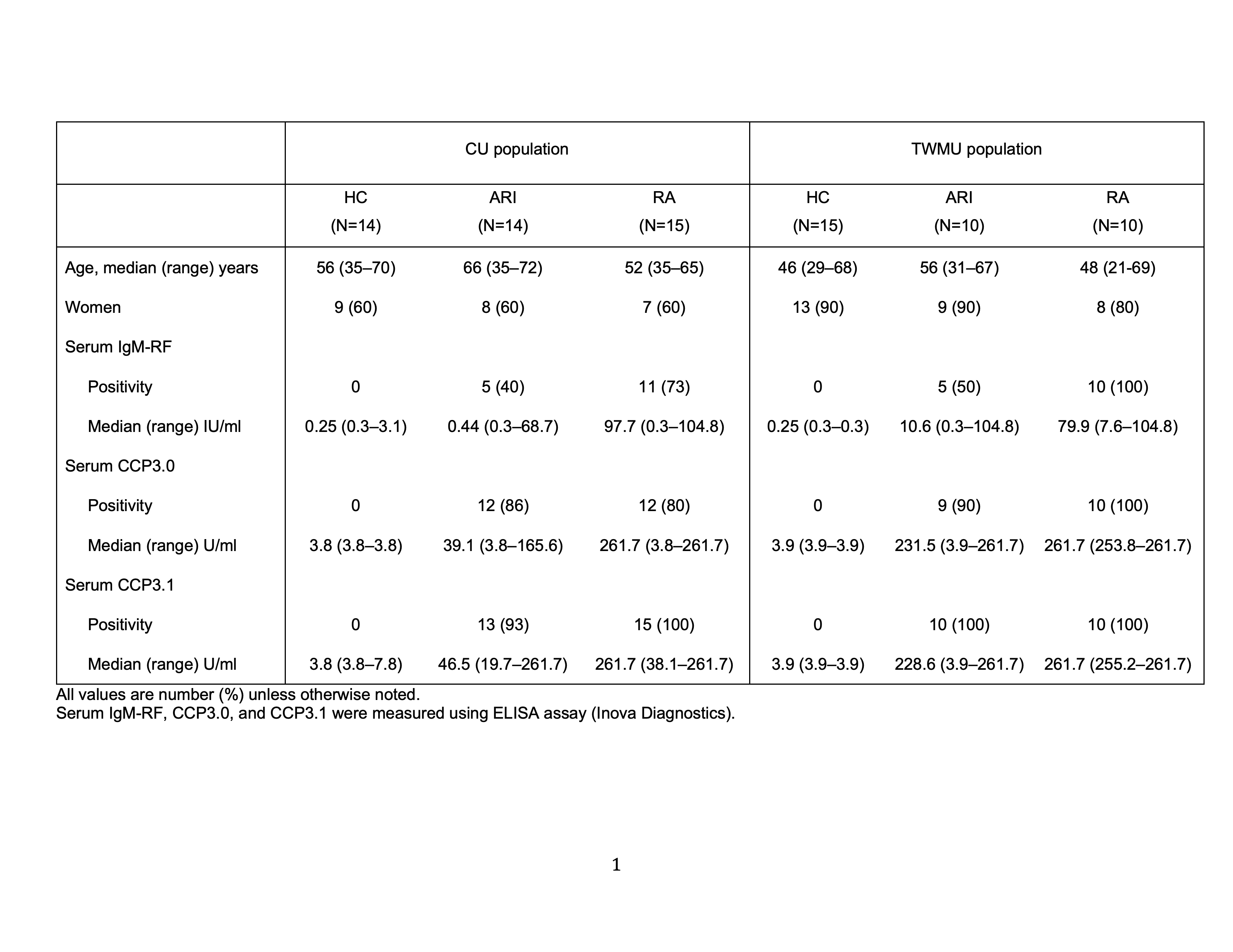
Methods: We recruited 14 and 10 ACPA+ individuals without arthritis but at-risk of future RA (ARI), 15 and 10 ACPA+ patients with early RA (2010 criteria and < 1 year from diagnosis), and 14 and 15 healthy controls (HC) from the Studies of the Etiologies of RA (SERA) population at University of Colorado (CU) and the rheumatology outpatient clinic at Tokyo Women’s Medical University (TWMU) in Japan, respectively (Table 1). Cryopreserved peripheral blood mononuclear cells were analyzed by flow cytometry to quantify cTph cells (PD-1hiCXCR5−CD4+ memory T cells) and cTfh cells (PD-1hiCXCR5+CD4+ memory T cells). We also assessed the expression of activation markers, HLA-DR and ICOS, on these cells.
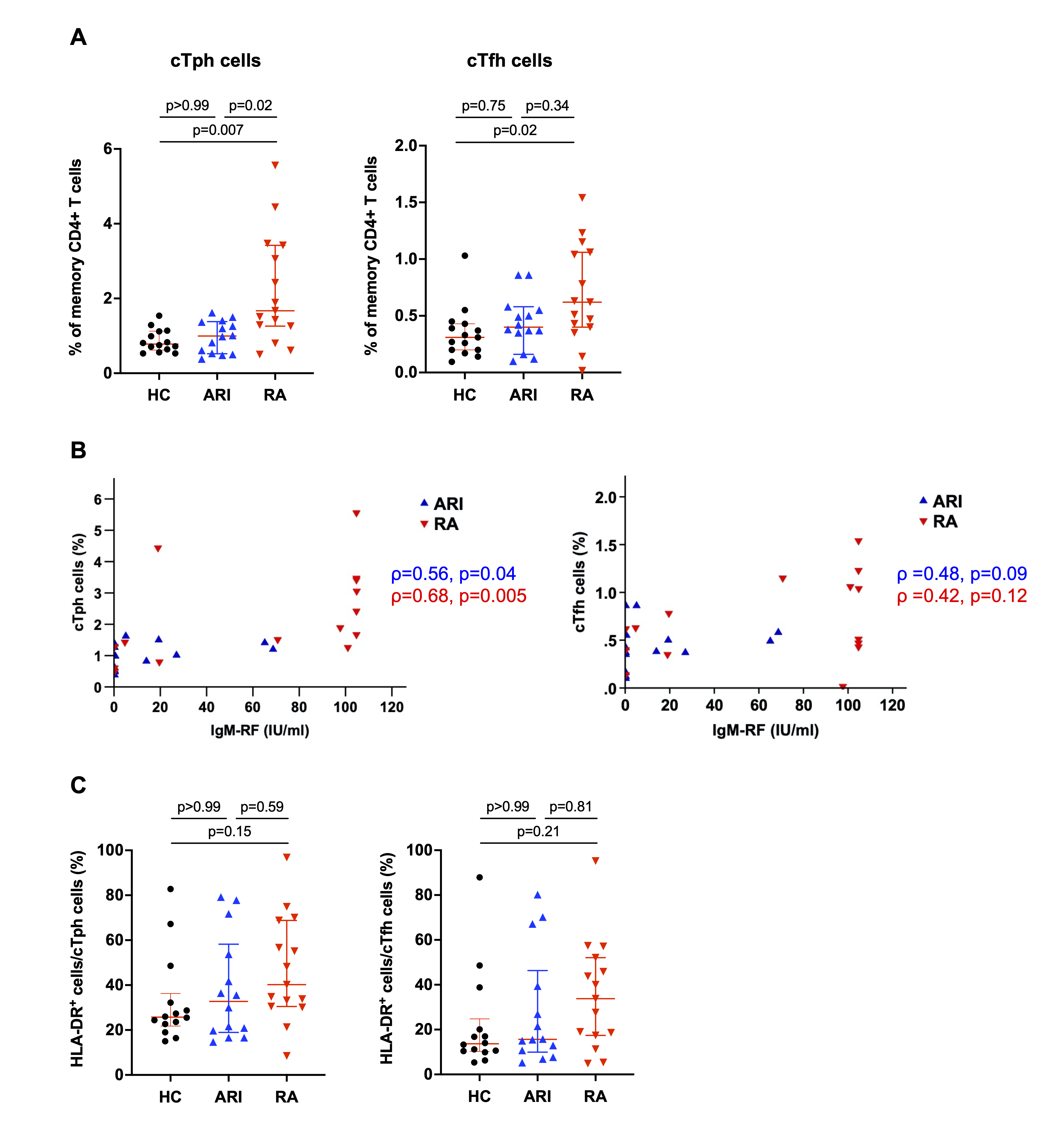
Results: In both ethnic populations, cTph cells were significantly increased in RA but not in ARI compared to HC (Figure 1A and 2A). In the CU population, the frequency of cTph cells was moderately correlated with serum IgM-RF levels in both ARI (Spearman’s ρ=0.56, p=0.04) and RA (Spearman’s ρ=0.68, p=0.005) (Figure 1B), while no correlation was observed between the frequencies of cTph cells and serum ACPA levels in either ARI or RA. There were no significant differences in the expressions of HLA-DR and ICOS on cTph cells or cTfh cells between CU study groups (Figure 1C). Unlike the CU population, the TWMU population did not show positive correlation between the frequency of cTph cells and serum IgM-RF levels in either ARI or RA (Figure 2B). In that population, however, the expression of HLA-DR on cTph cells was increased in both ARI and RA (Figure 2C).
Conclusion: cTph cells were expanded in RA but not in ARI in both ethnic populations, suggesting a relationship of these cells with clinically-apparent inflammatory arthritis. In addition, unique alterations in cTph cells among ACPA+ individuals with different ethnic backgrounds and in relationship to IgM-RF suggest their shared importance but potentially different roles in the future transition of ACPA+ ARI to early RA.
To cite this abstract in AMA style:
Takada H, Okamoto Y, Katsumata Y, Seifert J, Demoruelle K, Norris J, Deane K, Harigai M, Holers V. Unique Alterations in Circulating T Peripheral Helper Cells Are Found in Different Ethnic Groups of ACPA+ Individuals Both At-risk for and with Classified RA [abstract]. Arthritis Rheumatol. 2021; 73 (suppl 9). https://acrabstracts.org/abstract/unique-alterations-in-circulating-t-peripheral-helper-cells-are-found-in-different-ethnic-groups-of-acpa-individuals-both-at-risk-for-and-with-classified-ra/. Accessed .« Back to ACR Convergence 2021
ACR Meeting Abstracts - https://acrabstracts.org/abstract/unique-alterations-in-circulating-t-peripheral-helper-cells-are-found-in-different-ethnic-groups-of-acpa-individuals-both-at-risk-for-and-with-classified-ra/


