ACR Meeting Abstracts

ACR Meeting Abstracts

  • Meetings
    • ACR Convergence 2024
    • ACR Convergence 2023
    • 2023 ACR/ARP PRSYM
    • ACR Convergence 2022
    • ACR Convergence 2021
    • ACR Convergence 2020
    • 2020 ACR/ARP PRSYM
    • 2019 ACR/ARP Annual Meeting
    • 2018-2009 Meetings
    • Download Abstracts
  • Keyword Index
  • Advanced Search
  • Your Favorites
    • Favorites
    • Login
    • View and print all favorites
    • Clear all your favorites
  • ACR Meetings

Abstract Number: 0650

Tubulointerstitial Inflammation Predicts Tubular Atrophy, Fibrosis, and Renal Function Loss in Lupus Nephritis

Yevgeniya Gartshteyn1, Laura Geraldino2, Shuwei Wang3, Vivette D'Agati1 and Robert Winchester1, 1Columbia University Irving School of Medicine, New York, NY, 2Division of Rheumatology, Department of Medicine, Columbia University, New York, NY, 3Rutgers New Jersey Medical School, Newark, NJ

Meeting: ACR Convergence 2024

Keywords: Lupus nephritis, Nephritis, Systemic lupus erythematosus (SLE)

  • Tweet
  • Click to email a link to a friend (Opens in new window) Email
  • Click to print (Opens in new window) Print
Session Information

Date: Saturday, November 16, 2024

Title: SLE – Diagnosis, Manifestations, & Outcomes Poster I

Session Type: Poster Session A

Session Time: 10:30AM-12:30PM

Background/Purpose:     Current classification criteria of lupus nephritis (LN) emphasize glomerular pathology, however tubular atrophy and interstitial fibrosis (TAIF) are better predictors of poor renal outcomes.1 In this work, we evaluate tubulointerstitial inflammation (TII) as a risk factor for TAIF and renal failure.

Methods:     The subset of LN patients that underwent ≥2 renal biopsies, because of persistent or recurrent LN, at Columbia University Medical Center (CUMC) from 1994-2018 were identified. All renal biopsies were interpreted by nephropathologists at the CUMC Department of Pathology and classified using the ISN/RPS LN classification criteria. OPAL multiplex immunohistochemistry staining to identify CD3, CD4, CD8, CD19 and CD68 expression in the kidney tissue was performed.

Results:     104 LN patients (84% female, 25±12 years old at diagnosis, 14% White, 40% Black, 35% Hispanic) requiring sequential renal biopsies with a median follow-up of 11 [6-16] years were identified. On initial biopsy, 94/104 (90%) had class III or IV proliferative LN with or without class V.  Forty-seven (51%) patients developed ESRD. Nine patients died and 14 were lost to follow-up.
    Multivariable modeling of histopathological findings on biopsy 1 identified the presence of crescents to be associated with progression to ESRD (OR 3.14 95% CI 1.22, 8.08), while the presence of wire-loop lesions was protective (OR 0.28 98% CI 0.09, 0.85). On biopsy 2, the presence of TAIF≥25% (OR 4.50, 95% CI 1.30, 15.55) and glomerulosclerosis in >25% of glomeruli (OR 3.78, 95% CI 1.15, 0.03) were the only independent predictors of ESRD.
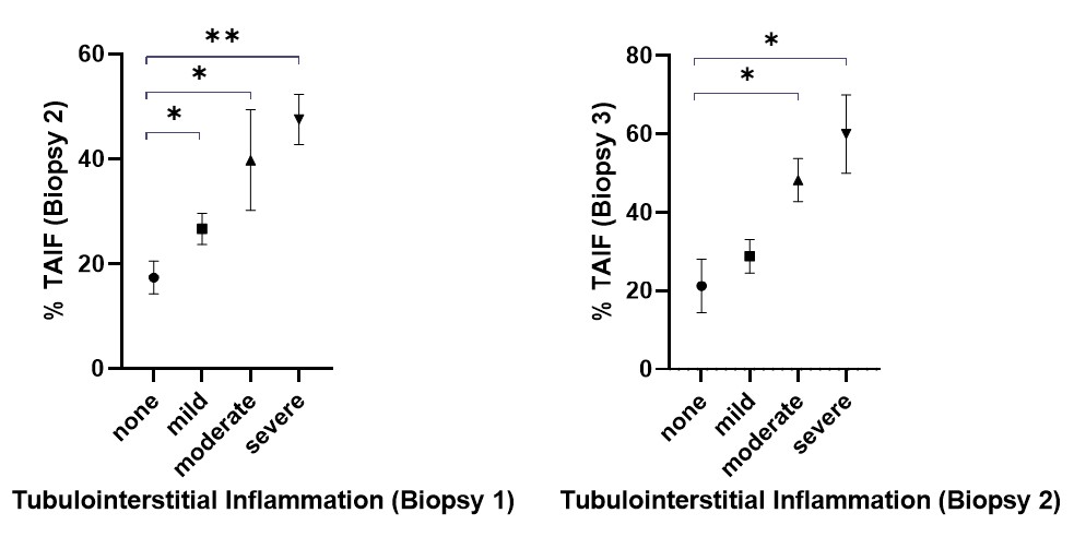
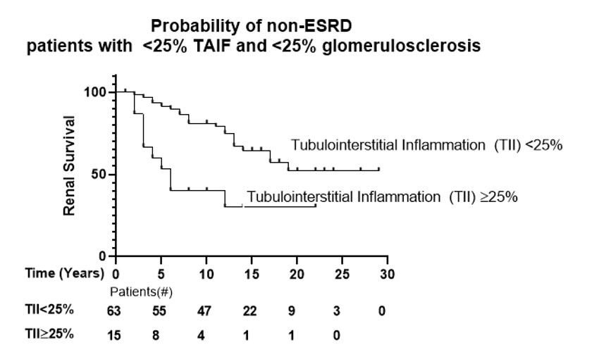
    There was a sequential increase in TAIF severity on successive biopsy samples (10%, 26%, 34%, and 46% TAIF on biopsy 1 through 4, respectively).  On univariable analysis, TAIF≥25% biopsy 2-4 predicted progression to ESRD (p< 0.01). The severity of TII on biopsy 1 was associated with increased risk of more advanced TAIF on biopsy 2, while TII on biopsy 2 predicted TAIF on biopsy 3, (Fig 1). Interestingly, in the 78 patients with < 25% TAIF and < 25% sclerotic glomerular involvement, moderate-severe TII significantly predicted ESRD (OR 4.76 95% CI 1.68, 13.50), (Fig 2).
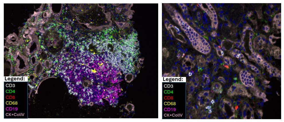
    Multiplex immunohistochemistry staining of kidney biopsy samples revealed that TII could be detected as organized immune infiltrate of T cell rich and B cell rich areas reminiscent of tertiary lymphoid structures (Fig 3, left) or as tubulitis mediated by CD3 T cells, often CD4neg and CD8neg (Fig 3, right).  

Conclusion:     TII predicts the development and extent of TAIF in lupus nephritis and is associated with a higher risk of progression to ESRD.

ACKNOWLEDGEMENT: The authors gratefully acknowledge the support of Dr. Anca D. Askanase and Dr. Adam Mor in this work.

1. Hsieh C et al.  Predicting outcomes of lupus nephritis with tubulointerstitial inflammation and scarring. Arthritis Care Res (Hoboken). 2011 Jun;63(6):865-74.

Supporting image 1

Figure 1. Severity of tubulointerstitial inflammation (TII) on biopsy 1 is associated with increased risk of more advanced TAIF on biopsy 2 (left). Severity of TII on biopsy 2 is associated with increased risk of more advanced TAIF on biopsy 3 (right).

Supporting image 2

Figure 2. In patients with <25% tubular atrophy and interstitial fibrosis (TAIF) and <25% of glomerulosclerosis on kidney biopsy, tubulointerstitial inflammation (TII) on the second biopsy predicts renal non-survival.

Supporting image 3

Figure 3. Confocal microscopy imaging shows examples of organized tubulointerstitial inflammation reminiscent of tertiary lymphoid structures with identifiable T-cell rich and B cell rich zones (left) as well as direct tubulitis by CD3 T cells (right). White, anti-CD3; Green, anti-CD4; Red, anti-CD8; Magenta, anti-CD19, Yellow, anti-CD68.


Disclosures: Y. Gartshteyn: None; L. Geraldino: Aurinia, 1, 2, Bristol Myers Squibb, 1, 2; S. Wang: None; V. D'Agati: None; R. Winchester: None.

To cite this abstract in AMA style:

Gartshteyn Y, Geraldino L, Wang S, D'Agati V, Winchester R. Tubulointerstitial Inflammation Predicts Tubular Atrophy, Fibrosis, and Renal Function Loss in Lupus Nephritis [abstract]. Arthritis Rheumatol. 2024; 76 (suppl 9). https://acrabstracts.org/abstract/tubulointerstitial-inflammation-predicts-tubular-atrophy-fibrosis-and-renal-function-loss-in-lupus-nephritis/. Accessed .
  • Tweet
  • Click to email a link to a friend (Opens in new window) Email
  • Click to print (Opens in new window) Print

« Back to ACR Convergence 2024

ACR Meeting Abstracts - https://acrabstracts.org/abstract/tubulointerstitial-inflammation-predicts-tubular-atrophy-fibrosis-and-renal-function-loss-in-lupus-nephritis/

Advanced Search

Your Favorites

You can save and print a list of your favorite abstracts during your browser session by clicking the “Favorite” button at the bottom of any abstract. View your favorites »

All abstracts accepted to ACR Convergence are under media embargo once the ACR has notified presenters of their abstract’s acceptance. They may be presented at other meetings or published as manuscripts after this time but should not be discussed in non-scholarly venues or outlets. The following embargo policies are strictly enforced by the ACR.

Accepted abstracts are made available to the public online in advance of the meeting and are published in a special online supplement of our scientific journal, Arthritis & Rheumatology. Information contained in those abstracts may not be released until the abstracts appear online. In an exception to the media embargo, academic institutions, private organizations, and companies with products whose value may be influenced by information contained in an abstract may issue a press release to coincide with the availability of an ACR abstract on the ACR website. However, the ACR continues to require that information that goes beyond that contained in the abstract (e.g., discussion of the abstract done as part of editorial news coverage) is under media embargo until 10:00 AM ET on November 14, 2024. Journalists with access to embargoed information cannot release articles or editorial news coverage before this time. Editorial news coverage is considered original articles/videos developed by employed journalists to report facts, commentary, and subject matter expert quotes in a narrative form using a variety of sources (e.g., research, announcements, press releases, events, etc.).

Violation of this policy may result in the abstract being withdrawn from the meeting and other measures deemed appropriate. Authors are responsible for notifying colleagues, institutions, communications firms, and all other stakeholders related to the development or promotion of the abstract about this policy. If you have questions about the ACR abstract embargo policy, please contact ACR abstracts staff at [email protected].

Wiley

  • Online Journal
  • Privacy Policy
  • Permissions Policies
  • Cookie Preferences

© Copyright 2025 American College of Rheumatology