ACR Meeting Abstracts

ACR Meeting Abstracts

  • Meetings
    • ACR Convergence 2024
    • ACR Convergence 2023
    • 2023 ACR/ARP PRSYM
    • ACR Convergence 2022
    • ACR Convergence 2021
    • ACR Convergence 2020
    • 2020 ACR/ARP PRSYM
    • 2019 ACR/ARP Annual Meeting
    • 2018-2009 Meetings
    • Download Abstracts
  • Keyword Index
  • Advanced Search
  • Your Favorites
    • Favorites
    • Login
    • View and print all favorites
    • Clear all your favorites
  • ACR Meetings

Abstract Number: 2899

Transcriptomic Meta-analysis of Lupus Affected Tissues Reveals Shared Immune, Metabolic, and Biochemical Dysregulation

Kathryn Kingsmore1, Sarah Heuer 1, Erika Hubbard 1, Michelle Catalina 2, Prathyusha Bachali 2, Peter Lipsky 3 and Amrie Grammer 1, 1AMPEL BioSolutions and RILITE Research Institute, Charlottesville, VA, 2AMPEL BioSolutions and the RILITE Research Institute, Charlottesville, VA, 3AMPEL BioSolutions, LLC, Charlottesville, VA

Meeting: 2019 ACR/ARP Annual Meeting

Keywords: discoid lupus erythematosus, lupus arthritis, Lupus nephritis, metabolism and bioinformatics

  • Tweet
  • Email
  • Print
Session Information

Date: Wednesday, November 13, 2019

Title: 6W022: SLE – Etiology & Pathogenesis II (2894–2899)

Session Type: ACR Abstract Session

Session Time: 11:00AM-12:30PM

Background/Purpose: Systemic lupus erythematosus (SLE) affects various organs and tissues, but whether pathologic processes in each organ are distinct or whether dysregulated molecular functions are found in common in all tissues is not known. We, therefore, conducted a meta-analysis of gene expression profiles in four affected SLE tissues to identify commonly dysregulated pathways.

Methods: Gene expression datasets for discoid lupus erythematosus (DLE), lupus arthritis (LA), lupus nephritis (LN) glomerulus (Glom), and LN tubulointerstitium (TI) were obtained from GEO. Differentially expressed genes (DEGs) were identified by LIMMA analysis for each dataset. DEGs from each tissue were analyzed with a multi-pronged bioinformatics approach to elucidate common immune cell infiltrates and common functional categories. These findings were then utilized to form modules of co-expressed genes to determine their enrichment using Gene Set Variation Analysis (GSVA).

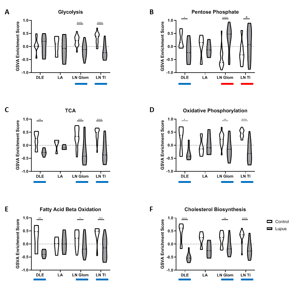
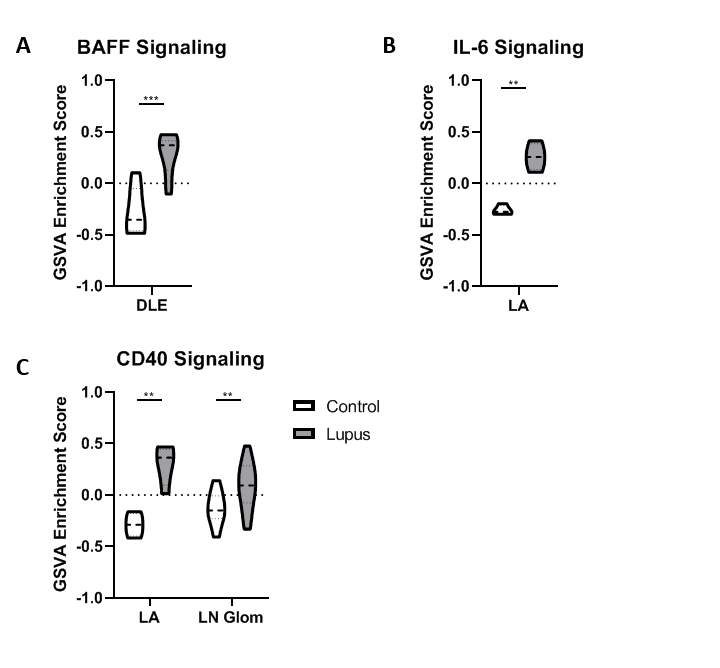
Results: All tissues demonstrated the presence of immune cells with the fewest immune cell transcripts in LN TI. Analysis of bulk gene expression revealed enrichment of antigen presenting cells (APCs), monocytes, and myeloid cells in all four tissues. Notably, enrichment of B cells, plasma cells, germinal center (GC) B cells, and CD8 T cells was only detected in DLE and LA. All four tissues demonstrated upregulated immune activity, including interferon-stimulated genes, pattern recognition receptors (PRRs), and antigen presentation (MHC Class II). Pro-apoptosis genes were also found enriched in DLE, LN Glom, and LN TI. A generalized decrease in biochemical processes was found in all four tissues and a specific decrease in both fatty acid biosynthesis and the tricarboxylic acid cycle was found in DLE and LN. Ingenuity Pathway Analysis (IPA®) further confirmed the activation of Dendritic Cell Maturation, Interferon, NFAT Regulation of Immune Response, PRRs, and TH1 signaling pathways in all four tissues. Additionally, IPA demonstrated cholesterol biosynthesis was decreased in all tissues except LA.

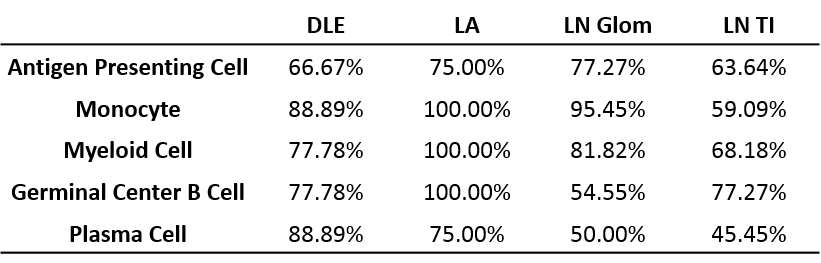
To confirm the aforementioned cellular infiltrates and aberrant pathways, as well as additional pathways, were operative in individual SLE tissues, GSVA was used to analyze enrichment of gene modules in patient samples. As shown in Table 1, Figure 1, and Figure 2, specific abnormalities were found in the majority of tissues, including enrichment of myeloid cells/monocytes, APCs, and GC B cells, whereas others were observed in some but not all tissues.

Conclusion: Common cellular infiltrates and molecular pathways were found in all affected tissues, suggesting commonalities in lupus organ pathogenesis. However, certain cell types and signaling were predominant in some tissues over others and GSVA illustrated heterogeneity between patients. Together this analysis informs a tissue-specific model of lupus immunopathogenesis and metabolic dysfunction with common and unique features and highlights the importance of patient specific identification of dysfunctional pathways in lupus organ pathogenesis.

Table 1 | Percentages of SLE Tissue Samples with GSVA enrichment of specific immune cell modules.

Figure 1 | GSVA demonstrates metabolic dysregulation in individual SLE affected tissues. GSVA enrichment scores calculated for -A- glycolysis, -B- pentose phosphate, -C- tricarboxylic acid cycle -TCA-, -D- oxidative phosphorylation, -E- fatty acid beta oxidation, and -F- cholesterol biosynthesis modules in DLE, LA, LN Glom and LN TI. Significant enrichment of tissue control to SLE affected tissue or SLE affected tissue to tissue control was determined using the Welch’s t-test. Red bar represents enrichment of SLE tissue over control and blue bar represents enrichment of tissue control over SLE tissue. # p < 0.1 *p < 0.05, ** p < 0.01, *** p < 0.001, **** < 0.0001.

Figure 2 | GSVA reveals potential pathways for therapeutic targeting in lupus affected tissues. Measures are shown for drug pathways significantly enriched in SLE affected tissue compared to control tissue as determined using the Welch’s t-test for -A- B cell activating factor -BAFF-, -B- interleukin -IL–6, and -C- CD40 signaling in DLE, LA, and LN Glom. ** p < 0.01, *** p < 0.001.


Disclosure: K. Kingsmore, None; S. Heuer, None; E. Hubbard, None; M. Catalina, None; P. Bachali, None; P. Lipsky, Horizon, 5, Janssen Research & Development, LLC, 2; A. Grammer, None.

To cite this abstract in AMA style:

Kingsmore K, Heuer S, Hubbard E, Catalina M, Bachali P, Lipsky P, Grammer A. Transcriptomic Meta-analysis of Lupus Affected Tissues Reveals Shared Immune, Metabolic, and Biochemical Dysregulation [abstract]. Arthritis Rheumatol. 2019; 71 (suppl 10). https://acrabstracts.org/abstract/transcriptomic-meta-analysis-of-lupus-affected-tissues-reveals-shared-immune-metabolic-and-biochemical-dysregulation/. Accessed .
  • Tweet
  • Email
  • Print

« Back to 2019 ACR/ARP Annual Meeting

ACR Meeting Abstracts - https://acrabstracts.org/abstract/transcriptomic-meta-analysis-of-lupus-affected-tissues-reveals-shared-immune-metabolic-and-biochemical-dysregulation/

Advanced Search

Your Favorites

You can save and print a list of your favorite abstracts during your browser session by clicking the “Favorite” button at the bottom of any abstract. View your favorites »

All abstracts accepted to ACR Convergence are under media embargo once the ACR has notified presenters of their abstract’s acceptance. They may be presented at other meetings or published as manuscripts after this time but should not be discussed in non-scholarly venues or outlets. The following embargo policies are strictly enforced by the ACR.

Accepted abstracts are made available to the public online in advance of the meeting and are published in a special online supplement of our scientific journal, Arthritis & Rheumatology. Information contained in those abstracts may not be released until the abstracts appear online. In an exception to the media embargo, academic institutions, private organizations, and companies with products whose value may be influenced by information contained in an abstract may issue a press release to coincide with the availability of an ACR abstract on the ACR website. However, the ACR continues to require that information that goes beyond that contained in the abstract (e.g., discussion of the abstract done as part of editorial news coverage) is under media embargo until 10:00 AM ET on November 14, 2024. Journalists with access to embargoed information cannot release articles or editorial news coverage before this time. Editorial news coverage is considered original articles/videos developed by employed journalists to report facts, commentary, and subject matter expert quotes in a narrative form using a variety of sources (e.g., research, announcements, press releases, events, etc.).

Violation of this policy may result in the abstract being withdrawn from the meeting and other measures deemed appropriate. Authors are responsible for notifying colleagues, institutions, communications firms, and all other stakeholders related to the development or promotion of the abstract about this policy. If you have questions about the ACR abstract embargo policy, please contact ACR abstracts staff at [email protected].

Wiley

  • Online Journal
  • Privacy Policy
  • Permissions Policies
  • Cookie Preferences

© Copyright 2025 American College of Rheumatology