Session Information
Date: Saturday, November 6, 2021
Title: Abstracts: Spondyloarthritis Including PsA – Diagnosis, Manifestations, & Outcomes I (0449–0452)
Session Type: Abstract Session
Session Time: 10:15AM-10:30AM
Background/Purpose: The underlying pathology in psoriatic arthritis (PsA) is systemic inflammation. The Total Body (TB)-PET/CT with the 18F-FDG radiotracer captures glucose metabolism across the entire body, and provides standardized measures as surrogates for degree of inflammation such as SUVmax. So, we predicted that these TB-PET/CT indices will correlate with the existing outcome measures of the 5 clinical domains of PsA (DAPSA, Leeds Enthesitis Index, Leeds Dactylitis Index, BASDAI and NAPSI). The objective of this study is to validate TB-PET/CT imaging as a diagnostic tool and for describing systemic disease activity of PsA.
Methods: We have prospectively recruited 40 participants (30 male, 10 female), with PsA (n=15), RA (n=10), and OA (n=15). All subjects underwent a single-timepoint TB-PET/CT scan on the uEXPLORER scanner using the PET radiotracer 18F-FDG. Qualitative findings and different patterns for these 3 conditions were evaluated. We quantified the degree of inflammation (SUVmax) and determined pathologic predilection for anatomical domains of bones/ligaments of in PsA, RA and OA.
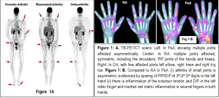
Results: In PsA patients, large number of joints showed positive findings in all participants. Multiple sites of enthesitis were visualized in the majority of scans (n=14/15). Furthermore, nail matrix showed increased uptake in 9/15 participants. Less frequent features included spine involvement of the supra/interspinous ligaments/bursae (n=6), sacroiliac joint (n=2), and dactylitis (n=2).
The uptake patterns and intensity were significantly different in patients with PsA compared to those with OA and RA (Fig 1 A and 1B). Fig 1 A, shows in PsA, joints were affected asymmetrically. In the Fig 1 B PET-CT imaging of the hands in PsA compared to RA clearly had: (a) asymmetry (b) also other characteristic pathologies such as in PsA there is inflammation of the extensor tendon and DIP in the left index along with marked nail matrix inflammation. The relative maximum standardized uptake value (rSUVmax) was significantly higher in participants with PsA compared to OA. There was a fair (68%) agreement between the DAPSA score and the PET averaged SUVmax. Figure 2 demonstrates the significance of a single TB-PET/CT scan in identifying the extent and the nature of joint, entheses, and nail involvement in PsA.
Conclusion: Our results indicates that TB-PET/CT measures can identify unique pathologies of PsA (Fig 1 and Fig 2) compared to RA and OA such as (i) asymmetric synovitis of large and small joints, (ii) systemic generalized enthesitis (iii) DIP inflammation, (iv) inflammation of the extensor tendons of fingers and its association with its enthesitis and adjacent nail matrix inflammation, (v) nail matrix inflammation, (vi) dactylitis, (vii) spondyloarthritis with sacroiliitis and diffuse enthesitis of the spine.
These observations substantiate our proposal that TB-PET/CT identifies the pathologies unique for 5 clinical domains of PsA, differentiates from RA/PsA, provides a diagnosis, extent/severity of the disease with a quantitative measure of total inflammatory burden and identifies subclinical developing pathologies of PsA. Thus, a diagnostic tool for early disease at the point of transition from psoriasis.

To cite this abstract in AMA style:
Raychaudhuri S, Abdelhafez Y, Kundu-Raychaudhuri S, Chaudhari A. Total-Body 18F-FDG PET/CT Imaging: A Tool for Diagnosis and Quantifying Inflammatory Burden of Psoriatic Arthritis [abstract]. Arthritis Rheumatol. 2021; 73 (suppl 9). https://acrabstracts.org/abstract/total-body-18f-fdg-pet-ct-imaging-a-tool-for-diagnosis-and-quantifying-inflammatory-burden-of-psoriatic-arthritis/. Accessed .« Back to ACR Convergence 2021
ACR Meeting Abstracts - https://acrabstracts.org/abstract/total-body-18f-fdg-pet-ct-imaging-a-tool-for-diagnosis-and-quantifying-inflammatory-burden-of-psoriatic-arthritis/