ACR Meeting Abstracts

ACR Meeting Abstracts

  • Meetings
    • ACR Convergence 2024
    • ACR Convergence 2023
    • 2023 ACR/ARP PRSYM
    • ACR Convergence 2022
    • ACR Convergence 2021
    • ACR Convergence 2020
    • 2020 ACR/ARP PRSYM
    • 2019 ACR/ARP Annual Meeting
    • 2018-2009 Meetings
    • Download Abstracts
  • Keyword Index
  • Advanced Search
  • Your Favorites
    • Favorites
    • Login
    • View and print all favorites
    • Clear all your favorites
  • ACR Meetings

Abstract Number: 1172

The Relationship Between Subclinical Inflammation and Bone Damage in Patients with Rheumatoid Arthritis Using Multimodality Imaging

Scott Brunet 1, Peter Salat 2, Glen Hazlewood 3, Klaus Engelke 4, Cheryl Barnabe 3 and Sarah Manske3, 1University of Calgary, Calgary, Canada, 2University of Calgary, Calary, Canada, 3University of Calgary, Calgary, AB, Canada, 4Erlangen University Hospital, Department of Medicine, Erlangen, Germany

Meeting: 2019 ACR/ARP Annual Meeting

Keywords: Imaging, magnetic resonance imaging (MRI) and computed tomography (CT), remission, Rheumatoid arthritis (RA)

  • Tweet
  • Email
  • Print
Session Information

Date: Monday, November 11, 2019

Title: Imaging Of Rheumatic Diseases Poster II

Session Type: Poster Session (Monday)

Session Time: 9:00AM-11:00AM

Background/Purpose: Many RA patients in clinical remission have evidence of bone marrow edema (BME) on magnetic resonance imaging (MRI), with radiographic bone damage progression appearing in a subset of patients.1 While MRI provides excellent visualization of subclinical inflammation, bone resolution is relatively poor, limiting the correlation of inflammatory imaging findings to erosive change that may develop. High resolution peripheral quantitative computed tomography (HR-pQCT) provides superior detailed bone resolution, with improved erosion detection and quantification. The purpose of this study was to determine whether subclinical inflammation observed on MRI could be co-localized with erosive change visualized by HR-pQCT, thereby predicting bone damage progression over a 6-month follow-up period for RA participants in clinical remission.

Methods: Nine participants meeting the ACR/EULAR criteria for RA and in clinical remission were recruited to undergo contrast-enhanced MRI and HR-pQCT scans of the hand at two timepoints, six months apart. Disease activity between these timepoints was assessed monthly by questionnaire, examination, and inflammatory biomarkers. A multimodal image registration technique was developed to co-localize imaging findings on HR-pQCT and MRI. Synovitis and BME were assessed semi-quantitatively with MRI using the RAMRIS scoring system.2 Bone erosion volume quantification was completed using a 3D segmentation technique using MIAF-Finger3, with joint space width (JSW) assessed using a volumetric approach from HR-pQCT images.4 Individual changes in HR-pQCT outcomes were compared against the least significant change (LSC) measured from scan-rescan reproducibility.

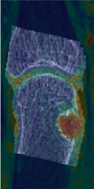
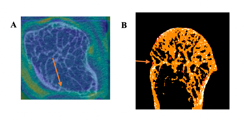
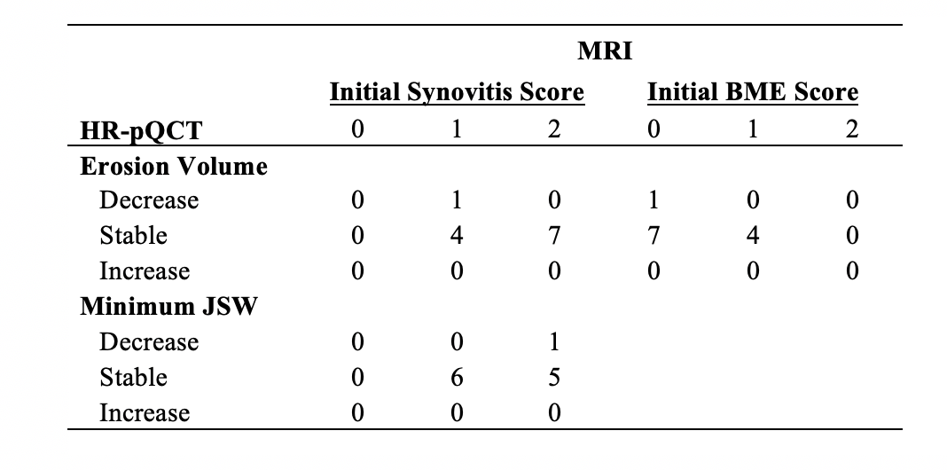
Results: The mean DAS28 for the 9 participants was 2.16 (SD 0.64) at the initial scan and 1.72 (SD 0.76) at follow-up. All participants had evidence of synovitis on MRI. Eight joints had evidence of BME at baseline. Twelve 2nd and 3rd MCP joints seen on HR-pQCT were co-localized with MRI using image registration (Figure 1). The presence of BME did not predict significant change in erosion volume (Table 1). In contrast, absence of BME and synovitis at the location of the erosion was associated with a decrease in erosion volume in one participant (Figure 2). The JSW outcomes remained stable in all joints, with the exception of one joint that showed a decrease in minimum JSW and another that had a decrease in the maximum JSW.

Conclusion: Even with highly sensitive imaging we did not detect erosive progression over the course of 6 months in patients with subclinical inflammation. However, the absence of localized subclinical inflammation was suggested to predict erosion healing. Findings will be confirmed in a larger cohort. This novel image registration technique for RA was able to co-localize areas of subclinical inflammation on MRI with joint damage on HR-pQCT, which could provide insights on the mechanisms of damage in RA that is not possible with either imaging approach alone.

References: 1. Brown, A. K. et al. Arthritis Rheum. 2008. 2. Østergaard, M. et al. Ann. Rheum. Dis. 2005. 3. Töpfer, D. et al. Rheumatol. (United Kingdom). 2014. 4. Stok, K. S. et al. J. Rheumatol.2017.

An HR-pQCT image overlaid with a transformed MRI image to localize inflammation -red- with bone damage.

-A- An erosion in HR-pQCT -bone = white- overlaid with MR image indicating no evidence of inflammation at the site of the erosion at baseline. -B- When baseline and follow-up HR-pQCT images are registered and aligned to visualize bone loss and gain, there is evidence of bone formation -red- at the erosion. This erosion showed a decrease in volume over 6 months. Orange indicates stable bone while white indicates resorbed bone.

Associations between MRI RAMRIS scores for synovitis and bone marrow edema -BME- at initial scan and 6-month changes in HR-pQCT erosion volume and minimum joint space width -JSW-. Decrease, Stable, and Increase characterization determined with respect to the Least Significant Change. -N = 12 joints from 7 participants-.


Disclosure: S. Brunet, None; P. Salat, None; G. Hazlewood, None; K. Engelke, None; C. Barnabe, None; S. Manske, None.

To cite this abstract in AMA style:

Brunet S, Salat P, Hazlewood G, Engelke K, Barnabe C, Manske S. The Relationship Between Subclinical Inflammation and Bone Damage in Patients with Rheumatoid Arthritis Using Multimodality Imaging [abstract]. Arthritis Rheumatol. 2019; 71 (suppl 10). https://acrabstracts.org/abstract/the-relationship-between-subclinical-inflammation-and-bone-damage-in-patients-with-rheumatoid-arthritis-using-multimodality-imaging/. Accessed .
  • Tweet
  • Email
  • Print

« Back to 2019 ACR/ARP Annual Meeting

ACR Meeting Abstracts - https://acrabstracts.org/abstract/the-relationship-between-subclinical-inflammation-and-bone-damage-in-patients-with-rheumatoid-arthritis-using-multimodality-imaging/

Advanced Search

Your Favorites

You can save and print a list of your favorite abstracts during your browser session by clicking the “Favorite” button at the bottom of any abstract. View your favorites »

All abstracts accepted to ACR Convergence are under media embargo once the ACR has notified presenters of their abstract’s acceptance. They may be presented at other meetings or published as manuscripts after this time but should not be discussed in non-scholarly venues or outlets. The following embargo policies are strictly enforced by the ACR.

Accepted abstracts are made available to the public online in advance of the meeting and are published in a special online supplement of our scientific journal, Arthritis & Rheumatology. Information contained in those abstracts may not be released until the abstracts appear online. In an exception to the media embargo, academic institutions, private organizations, and companies with products whose value may be influenced by information contained in an abstract may issue a press release to coincide with the availability of an ACR abstract on the ACR website. However, the ACR continues to require that information that goes beyond that contained in the abstract (e.g., discussion of the abstract done as part of editorial news coverage) is under media embargo until 10:00 AM ET on November 14, 2024. Journalists with access to embargoed information cannot release articles or editorial news coverage before this time. Editorial news coverage is considered original articles/videos developed by employed journalists to report facts, commentary, and subject matter expert quotes in a narrative form using a variety of sources (e.g., research, announcements, press releases, events, etc.).

Violation of this policy may result in the abstract being withdrawn from the meeting and other measures deemed appropriate. Authors are responsible for notifying colleagues, institutions, communications firms, and all other stakeholders related to the development or promotion of the abstract about this policy. If you have questions about the ACR abstract embargo policy, please contact ACR abstracts staff at [email protected].

Wiley

  • Online Journal
  • Privacy Policy
  • Permissions Policies
  • Cookie Preferences

© Copyright 2025 American College of Rheumatology